|
|
|
|
 Description
|
|
Compounds
|
||||||||||||||||||||||||||||||||||||||||||||||||||||
Chains, Units
Summary Information (see also Sequences/Alignments below) |
Ligands, Modified Residues, Ions (0, 0)| (no "Ligand,Modified Residues,Ions" information available for 1W4J) |
Sites (0, 0)| (no "Site" information available for 1W4J) |
SS Bonds (0, 0)| (no "SS Bond" information available for 1W4J) |
Cis Peptide Bonds (0, 0)| (no "Cis Peptide Bond" information available for 1W4J) |
SAPs(SNPs)/Variants (0, 0)| (no "SAP(SNP)/Variant" information available for 1W4J) |
PROSITE Motifs (0, 0)| (no "PROSITE Motif" information available for 1W4J) |
Exons (0, 0)| (no "Exon" information available for 1W4J) |
Sequences/Alignments
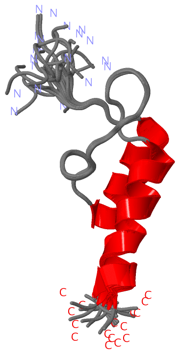
NMR StructureChain A from PDB Type:PROTEIN Length:51 aligned with Q8ZUR6_PYRAE | Q8ZUR6 from UniProtKB/TrEMBL Length:383 Alignment length:59 92 102 112 122 132 Q8ZUR6_PYRAE 83 GPQTEAPARPREVAAMPAARRLAKELGIDLSKVKGTGPGGVITVEDVKRYAEETAKATA 141 SCOP domains ----------------------------------------------------------- SCOP domains CATH domains 1 w4jA00 A:125-175 Dihydrolipoamide Transferase CATH domains Pfam domains ----------E3_binding-1w4jA01 A:127-165 ---------- Pfam domains
|
||||||||||||||||||||
SCOP Domains (0, 0)| (no "SCOP Domain" information available for 1W4J) |
CATH Domains (1, 1)
NMR Structure
 |
Pfam Domains (1, 1)
NMR Structure
|
Gene Ontology (3, 3)|
NMR Structure(hide GO term definitions) Chain A (Q8ZUR6_PYRAE | Q8ZUR6)
|
||||||||||||||||||||||||||||||
Interactive Views
|
||||||||||||||||||||||||||||||||||||||||||||||||||||||||||||||||||||||||||||||||||||||||||||||||||||||||||||||||||||
Still Images
|
||||||||||||||||
Databases
|
||||||||||||||||||||||||||||||||||||||||||||||||||||||||||||||||||||||||||||||||||||||||||||||||||||||||||||||||||||||||||||||||||||||||||||||||||||||||||||||||
Analysis Tools
|
|||||||||||||||||||||||||||||||||||||||||||||||||||||||||||||
Entries Sharing at Least One Protein Chain (UniProt ID)
Related Entries Specified in the PDB File
|
|