Show PDB file:   
         Plain Text   HTML   (compressed file size)
QuickSearch:   
by PDB,NDB,UniProt,PROSITE Code or Search Term(s)  
(-)Asym./Biol. Unit
(-)Asym./Biol. Unit - sites
collapse expand < >
Image Asym./Biol. Unit
Asym./Biol. Unit  (Jmol Viewer)
Image Asym./Biol. Unit - sites
Asym./Biol. Unit - sites  (Jmol Viewer)

(-) Description

Title :  CYTOCHROME C DOMAIN OF CAA3 OXYGEN OXIDOREDUCTASE
 
Authors :  V. Srinivasan, C. Rajendran, F. L. Sousa, A. M. P. Melo, L. M. Saraiva, M. M. Pereira, M. Santana, M. Teixeira, H. Michel
Date :  06 Jul 04  (Deposition) - 19 Jan 05  (Release) - 24 Feb 09  (Revision)
Method :  X-RAY DIFFRACTION
Resolution :  1.30
Chains :  Asym./Biol. Unit :  A
Keywords :  Cytochrome C Domain, Oxidoreductase (Keyword Search: [Gene Ontology, PubMed, Web (Google))
 
Reference :  V. Srinivasan, C. Rajendran, F. L. Sousa, A. M. P. Melo, L. M. Saraiva, M. M. Pereira, M. Santana, M. Teixeira, H. Michel
Structure At 1. 3A Resolution Of Rhodothermus Marinus Caa(3) Cytochrome C Domain
J. Mol. Biol. V. 345 1047 2005
PubMed-ID: 15644203  |  Reference-DOI: 10.1016/J.JMB.2004.10.069

(-) Compounds

Molecule 1 - CYTOCHROME OXIDASE SUBUNIT II
    ChainsA
    EC Number1.9.3.1
    EngineeredYES
    Expression SystemESCHERICHIA COLI
    Expression System StrainBL21
    Expression System Taxid511693
    Expression System VectorPET-12A
    FragmentCYTOCHROME C DOMAIN, RESIDUES 218-316
    Organism ScientificRHODOTHERMUS MARINUS
    Organism Taxid29549

 Structural Features

(-) Chains, Units

  1
Asymmetric/Biological Unit A

Summary Information (see also Sequences/Alignments below)
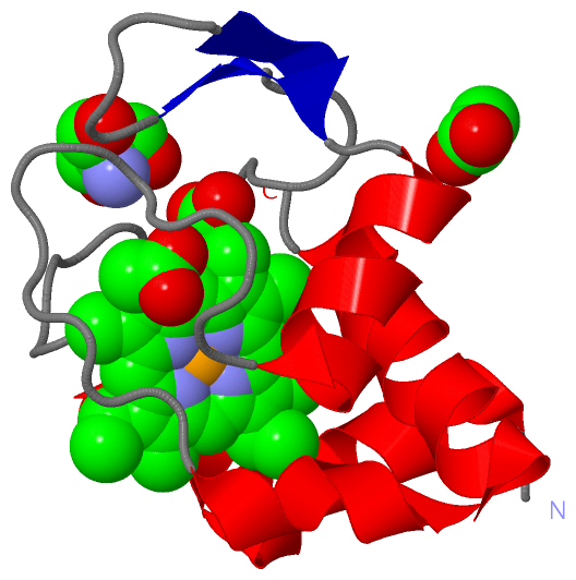
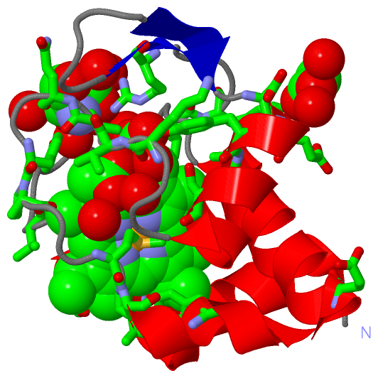
(-) Ligands, Modified Residues, Ions  (3, 3)

Asymmetric/Biological Unit (3, 3)
No.NameCountTypeFull Name
1ACT1Ligand/IonACETATE ION
2HEM1Ligand/IonPROTOPORPHYRIN IX CONTAINING FE
3TRS1Ligand/Ion2-AMINO-2-HYDROXYMETHYL-PROPANE-1,3-DIOL

(-) Sites  (3, 3)

Asymmetric Unit (3, 3)
No.NameEvidenceResiduesDescription
1AC1SOFTWAREALA A:17 , SER A:20 , TYR A:38 , ASP A:53 , GLU A:54 , PRO A:73 , HOH A:2130 , HOH A:2131BINDING SITE FOR RESIDUE ACT A1102
2AC2SOFTWAREALA A:17 , CYS A:18 , CYS A:21 , HIS A:22 , VAL A:30 , PRO A:32 , LEU A:37 , ARG A:42 , TYR A:56 , LEU A:57 , SER A:60 , LYS A:67 , VAL A:68 , VAL A:69 , PRO A:73 , MET A:76 , PRO A:77 , TYR A:80 , HOH A:2046 , HOH A:2124 , HOH A:2125 , HOH A:2126BINDING SITE FOR RESIDUE HEM A1100
3AC3SOFTWAREARG A:42 , VAL A:69 , GLN A:70 , TYR A:72 , GLU A:85 , HOH A:2044 , HOH A:2058 , HOH A:2127 , HOH A:2128 , HOH A:2129BINDING SITE FOR RESIDUE TRS A1101

(-) SS Bonds  (0, 0)

(no "SS Bond" information available for 1W2L)

(-) Cis Peptide Bonds  (0, 0)

(no "Cis Peptide Bond" information available for 1W2L)

 Sequence-Structure Mapping

(-) SAPs(SNPs)/Variants  (0, 0)

(no "SAP(SNP)/Variant" information available for 1W2L)

(-) PROSITE Motifs  (0, 0)

(no "PROSITE Motif" information available for 1W2L)

(-) Exons   (0, 0)

(no "Exon" information available for 1W2L)

(-) Sequences/Alignments

Asymmetric/Biological Unit
   Reformat: Number of residues per line =  ('0' or empty: single-line sequence representation)
  Number of residues per labelling interval =   
  UniProt sequence: complete  aligned part    
   Show mapping: SCOP domains CATH domains Pfam domains Secondary structure (by author)
SAPs(SNPs) PROSITE motifs Exons
(details for a mapped element are shown in a popup box when the mouse pointer rests over it)
Chain A from PDB  Type:PROTEIN  Length:97
 aligned with Q9F3S9_RHOMR | Q9F3S9 from UniProtKB/TrEMBL  Length:316

    Alignment length:97
                                   229       239       249       259       269       279       289       299       309       
         Q9F3S9_RHOMR   220 MPLAELGARLYREKACFSCHSIDGSRLVGPSFKGLYGSTRTFEDGTTAVADENYLRESILQPGAKVVQGYPNVMPASYASLSEREVAALIEFIKQQQ 316
               SCOP domains d1w2la_ A: automated matches                                                                      SCOP domains
               CATH domains ------------------------------------------------------------------------------------------------- CATH domains
               Pfam domains -Cytochrome_CBB3-1w2lA01 A:4-95                                                              ---- Pfam domains
         Sec.struct. author .hhhhhhhhhhhhhhhhhh...................eee.....eee.hhhhhhhhhhh.............hhhhhhhhhhhhhhhhhhhhh.. Sec.struct. author
                 SAPs(SNPs) ------------------------------------------------------------------------------------------------- SAPs(SNPs)
                    PROSITE ------------------------------------------------------------------------------------------------- PROSITE
                 Transcript ------------------------------------------------------------------------------------------------- Transcript
                 1w2l A   3 MPLAELGARLYREKACFSCHSIDGSRLVGPSFKGLYGSTRTFEDGTTAVADENYLRESILQPGAKVVQGYPNVMPASYASLSEREVAALIEFIKQQQ  99
                                    12        22        32        42        52        62        72        82        92       

   Legend:   → Mismatch (orange background)
  - → Gap (green background, '-', border residues have a numbering label)
    → Modified Residue (blue background, lower-case, 'x' indicates undefined single-letter code, labelled with number + name)
  x → Chemical Group (purple background, 'x', labelled with number + name, e.g. ACE or NH2)
  extra numbering lines below/above indicate numbering irregularities and modified residue names etc., number ends below/above '|'

 Classification and Annotation

(-) SCOP Domains  (1, 1)

Asymmetric/Biological Unit

(-) CATH Domains  (0, 0)

(no "CATH Domain" information available for 1W2L)

(-) Pfam Domains  (1, 1)

Asymmetric/Biological Unit

(-) Gene Ontology  (13, 13)

Asymmetric/Biological Unit(hide GO term definitions)
Chain A   (Q9F3S9_RHOMR | Q9F3S9)
molecular function
    GO:0005507    copper ion binding    Interacting selectively and non-covalently with copper (Cu) ions.
    GO:0004129    cytochrome-c oxidase activity    Catalysis of the reaction: 4 ferrocytochrome c + O2 + 4 H+ = 4 ferricytochrome c + 2 H2O.
    GO:0009055    electron carrier activity    Any molecular entity that serves as an electron acceptor and electron donor in an electron transport chain. An electron transport chain is a process in which a series of electron carriers operate together to transfer electrons from donors to any of several different terminal electron acceptors to generate a transmembrane electrochemical gradient.
    GO:0020037    heme binding    Interacting selectively and non-covalently with heme, any compound of iron complexed in a porphyrin (tetrapyrrole) ring.
    GO:0046872    metal ion binding    Interacting selectively and non-covalently with any metal ion.
    GO:0016491    oxidoreductase activity    Catalysis of an oxidation-reduction (redox) reaction, a reversible chemical reaction in which the oxidation state of an atom or atoms within a molecule is altered. One substrate acts as a hydrogen or electron donor and becomes oxidized, while the other acts as hydrogen or electron acceptor and becomes reduced.
biological process
    GO:0022900    electron transport chain    A process in which a series of electron carriers operate together to transfer electrons from donors to any of several different terminal electron acceptors to generate a transmembrane electrochemical gradient.
    GO:1902600    hydrogen ion transmembrane transport    The directed movement of hydrogen ion (proton) across a membrane.
    GO:0055114    oxidation-reduction process    A metabolic process that results in the removal or addition of one or more electrons to or from a substance, with or without the concomitant removal or addition of a proton or protons.
cellular component
    GO:0016021    integral component of membrane    The component of a membrane consisting of the gene products and protein complexes having at least some part of their peptide sequence embedded in the hydrophobic region of the membrane.
    GO:0016020    membrane    A lipid bilayer along with all the proteins and protein complexes embedded in it an attached to it.
    GO:0005886    plasma membrane    The membrane surrounding a cell that separates the cell from its external environment. It consists of a phospholipid bilayer and associated proteins.
    GO:0070469    respiratory chain    The protein complexes that form the electron transport system (the respiratory chain), associated with a cell membrane, usually the plasma membrane (in prokaryotes) or the inner mitochondrial membrane (on eukaryotes). The respiratory chain complexes transfer electrons from an electron donor to an electron acceptor and are associated with a proton pump to create a transmembrane electrochemical gradient.

 Visualization

(-) Interactive Views

Asymmetric/Biological Unit
  Complete Structure
    Jena3D(integrated viewing of ligand, site, SAP, PROSITE, SCOP information)
    WebMol | AstexViewer[tm]@PDBe
(Java Applets, require no local installation except for Java; loading may be slow)
    STRAP
(Java WebStart application, automatic local installation, requires Java; full application with system access!)
    RasMol
(require local installation)
    Molscript (VRML)
(requires installation of a VRML viewer; select preferred view via VRML and generate a mono or stereo PDF format file)
 
  Ligands, Modified Residues, Ions
    ACT  [ RasMol | Jena3D ]  +environment [ RasMol | Jena3D ]
    HEM  [ RasMol | Jena3D ]  +environment [ RasMol | Jena3D ]
    TRS  [ RasMol | Jena3D ]  +environment [ RasMol | Jena3D ]
 
  Sites
    AC1  [ RasMol ]  +environment [ RasMol ]
    AC2  [ RasMol ]  +environment [ RasMol ]
    AC3  [ RasMol ]  +environment [ RasMol ]
 
  Cis Peptide Bonds
(no "Cis Peptide Bonds" information available for 1w2l)
 

(-) Still Images

Jmol
  protein: cartoon or spacefill or dots and stick; nucleic acid: cartoon and stick; ligands: spacefill; active site: stick
Molscript
  protein, nucleic acid: cartoon; ligands: spacefill; active site: ball and stick

 Databases and Analysis Tools

(-) Databases

Access by PDB/NDB ID
  1w2l
    Family and Domain InformationProDom | SYSTERS
    General Structural InformationGlycoscienceDB | MMDB | NDB | OCA | PDB | PDBe | PDBj | PDBsum | PDBWiki | PQS | PROTEOPEDIA
    Orientation in MembranesOPM
    Protein SurfaceSURFACE
    Secondary StructureDSSP (structure derived) | HSSP (homology derived)
    Structural GenomicsGeneCensus
    Structural NeighboursCE | VAST
    Structure ClassificationCATH | Dali | SCOP
    Validation and Original DataBMRB Data View | BMRB Restraints Grid | EDS | PROCHECK | RECOORD | WHAT_CHECK
 
Access by UniProt ID/Accession number
  Q9F3S9_RHOMR | Q9F3S9
    Comparative Protein Structure ModelsModBase
    Genomic InformationEnsembl
    Protein-protein InteractionDIP
    Sequence, Family and Domain InformationInterPro | Pfam | SMART | UniProtKB/TrEMBL
 
Access by Enzyme Classificator   (EC Number)
  1.9.3.1
    General Enzyme InformationBRENDA | EC-PDB | Enzyme | IntEnz
    PathwayKEGG | MetaCyc
 
Access by Disease Identifier   (MIM ID)
  (no 'MIM ID' available)
    Disease InformationOMIM
 
Access by GenAge ID
  (no 'GenAge ID' available)
    Age Related InformationGenAge

(-) Analysis Tools

Access by PDB/NDB ID
    Domain InformationXDom
    Interatomic Contacts of Structural UnitsCSU
    Ligand-protein ContactsLPC
    Protein CavitiescastP
    Sequence and Secondary StructurePDBCartoon
    Structure AlignmentSTRAP(Java WebStart application, automatic local installation, requires Java; full application with system access!)
    Structure and Sequence BrowserSTING
 
Access by UniProt ID/Accession number
  Q9F3S9_RHOMR | Q9F3S9
    Protein Disorder PredictionDisEMBL | FoldIndex | GLOBPLOT (for more information see DisProt)

 Related Entries

(-) Entries Sharing at Least One Protein Chain (UniProt ID)

(no "Entries Sharing at Least One Protein Chain" available for 1W2L)

(-) Related Entries Specified in the PDB File

(no "Related Entries Specified in the PDB File" available for 1W2L)