| molecular function |
|---|
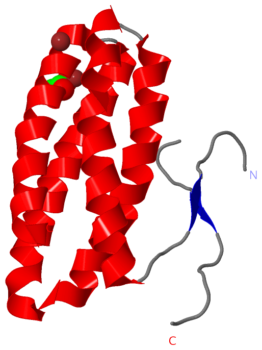
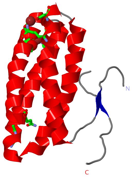
| | GO:0005509 | | calcium ion binding | | Interacting selectively and non-covalently with calcium ions (Ca2+). |
| biological process |
|---|
| | GO:0015979 | | photosynthesis | | The synthesis by organisms of organic chemical compounds, especially carbohydrates, from carbon dioxide (CO2) using energy obtained from light rather than from the oxidation of chemical compounds. |
| cellular component |
|---|
| | GO:0009507 | | chloroplast | | A chlorophyll-containing plastid with thylakoids organized into grana and frets, or stroma thylakoids, and embedded in a stroma. |
| | GO:0009535 | | chloroplast thylakoid membrane | | The pigmented membrane of a chloroplast thylakoid. An example of this component is found in Arabidopsis thaliana. |
| | GO:0019898 | | extrinsic component of membrane | | The component of a membrane consisting of gene products and protein complexes that are loosely bound to one of its surfaces, but not integrated into the hydrophobic region. |
| | GO:0016020 | | membrane | | A lipid bilayer along with all the proteins and protein complexes embedded in it an attached to it. |
| | GO:0009523 | | photosystem II | | A photosystem that contains a pheophytin-quinone reaction center with associated accessory pigments and electron carriers. In cyanobacteria and chloroplasts, in the presence of light, PSII functions as a water-plastoquinone oxidoreductase, transferring electrons from water to plastoquinone, whereas other photosynthetic bacteria carry out anoxygenic photosynthesis and oxidize other compounds to re-reduce the photoreaction center. |
| | GO:0009654 | | photosystem II oxygen evolving complex | | A complex, composed of a cluster of manganese, calcium and chloride ions bound to extrinsic proteins, that catalyzes the splitting of water to O2 and 4 H+. In cyanobacteria there are five extrinsic proteins in OEC (PsbO, PsbP-like, PsbQ-like, PsbU and PsbV), while in plants there are only three (PsbO, PsbP and PsbQ). |
| | GO:0009536 | | plastid | | Any member of a family of organelles found in the cytoplasm of plants and some protists, which are membrane-bounded and contain DNA. Plant plastids develop from a common type, the proplastid. |
| | GO:0009579 | | thylakoid | | A membranous cellular structure that bears the photosynthetic pigments in plants, algae, and cyanobacteria. In cyanobacteria thylakoids are of various shapes and are attached to, or continuous with, the plasma membrane. In eukaryotes they are flattened, membrane-bounded disk-like structures located in the chloroplasts; in the chloroplasts of higher plants the thylakoids form dense stacks called grana. Isolated thylakoid preparations can carry out photosynthetic electron transport and the associated phosphorylation. |
 Description