|
|
|
|
 Description
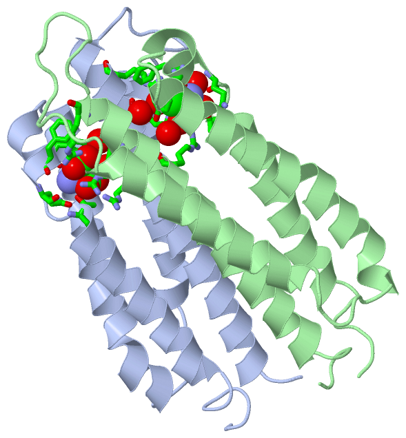
|
|
Compounds
|
||||||||||||||||||||||||
Chains, Units
Summary Information (see also Sequences/Alignments below) |
Ligands, Modified Residues, Ions (1, 2)
Asymmetric/Biological Unit (1, 2)
|
Sites (2, 2)
Asymmetric Unit (2, 2)
|
SS Bonds (0, 0)| (no "SS Bond" information available for 1VLT) |
Cis Peptide Bonds (0, 0)| (no "Cis Peptide Bond" information available for 1VLT) |
SAPs(SNPs)/Variants (0, 0)| (no "SAP(SNP)/Variant" information available for 1VLT) |
PROSITE Motifs (0, 0)| (no "PROSITE Motif" information available for 1VLT) |
Exons (0, 0)| (no "Exon" information available for 1VLT) |
Sequences/Alignments
Asymmetric/Biological UnitChain A from PDB Type:PROTEIN Length:143 aligned with MCP2_SALTY | P02941 from UniProtKB/Swiss-Prot Length:553 Alignment length:144 48 58 68 78 88 98 108 118 128 138 148 158 168 178 MCP2_SALTY 39 GFVISNELRQQQSELTSTWDLMLQTRINLSRSAARMMMDASNQQSSAKTDLLQNAKTTLAQAAAHYANFKNMTPLPAMAEASANVDEKYQRYQAALAELIQFLDNGNMDAYFAQPTQGMQNALGEALGNYARVSENLYRQTFDQ 182 SCOP domains d1vlta_ A: Aspartate receptor, ligand-binding domain SCOP domains CATH domains 1vltA00 A:39-180 Aspartate receptor Chemotaxis Sensory Transducers Family -- CATH domains Pfam domains ------------------------------------------------------------------------------------------------------------------------------------------------ Pfam domains SAPs(SNPs) ------------------------------------------------------------------------------------------------------------------------------------------------ SAPs(SNPs) PROSITE ------------------------------------------------------------------------------------------------------------------------------------------------ PROSITE Transcript ------------------------------------------------------------------------------------------------------------------------------------------------ Transcript 1vlt A 39 GFVISNELRQQQSELTSTWDLMLQTRINLSRSAARMMMDASNQQSSAKTDLLQNAKTTLAQAAAHYANFKNMTPLPAMAEASANVDEKYQRYQAALAELIQFLDNGNMDAYFAQPTQGMQNALGEALGNYARVSENLYRQTF-D 389 48 58 68 78 88 98 108 118 128 138 148 158 168 178 | | 180 | 389 Chain B from PDB Type:PROTEIN Length:144 aligned with MCP2_SALTY | P02941 from UniProtKB/Swiss-Prot Length:553 Alignment length:145 47 57 67 77 87 97 107 117 127 137 147 157 167 177 MCP2_SALTY 38 QGFVISNELRQQQSELTSTWDLMLQTRINLSRSAARMMMDASNQQSSAKTDLLQNAKTTLAQAAAHYANFKNMTPLPAMAEASANVDEKYQRYQAALAELIQFLDNGNMDAYFAQPTQGMQNALGEALGNYARVSENLYRQTFDQ 182 SCOP domains d1vltb_ B: Aspartate receptor, ligand-binding domain SCOP domains CATH domains 1vltB00 B:38-180 Aspartate receptor Chemotaxis Sensory Transducers Family -- CATH domains Pfam domains ------------------------------------------------------------------------------------------------------------------------------------------------- Pfam domains SAPs(SNPs) ------------------------------------------------------------------------------------------------------------------------------------------------- SAPs(SNPs) PROSITE ------------------------------------------------------------------------------------------------------------------------------------------------- PROSITE Transcript ------------------------------------------------------------------------------------------------------------------------------------------------- Transcript 1vlt B 38 QGFVISNELRQQQSELTSTWDLMLQTRINLSRSAARMMMDASNQQSSAKTDLLQNAKTTLAQAAAHYANFKNMTPLPAMAEASANVDEKYQRYQAALAELIQFLDNGNMDAYFAQPTQGMQNALGEALGNYARVSENLYRQTF-D 189 47 57 67 77 87 97 107 117 127 137 147 157 167 177 | | 180 | 189
|
||||||||||||||||||||
SCOP Domains (1, 2)
Asymmetric/Biological Unit
 |
CATH Domains (1, 2)
Asymmetric/Biological Unit
|
Pfam Domains (0, 0)| (no "Pfam Domain" information available for 1VLT) |
Gene Ontology (7, 7)|
Asymmetric/Biological Unit(hide GO term definitions) Chain A,B (MCP2_SALTY | P02941)
|
||||||||||||||||||||||||||||||||||||||||||||||||||||||||||||
Interactive Views
|
|||||||||||||||||||||||||||||||||||||||||||||||||||||||||||||||||||||||||||||||||||||||||||||||||||||||||||||||||||||||||||||
Still Images
|
||||||||||||||||
Databases
|
||||||||||||||||||||||||||||||||||||||||||||||||||||||||||||||||||||||||||||||||||||||||||||||||||||||||||||||||||||||||||||||||||||||||||||||||||||||||||||||||
Analysis Tools
|
|||||||||||||||||||||||||||||||||||||||||||||||||||||||||||||
Entries Sharing at Least One Protein Chain (UniProt ID)
Related Entries Specified in the PDB File
|
|