|
|
|
|
 Description
|
|
Compounds
|
||||||||||||||||||||||||||||
Chains, Units
Summary Information (see also Sequences/Alignments below) |
Ligands, Modified Residues, Ions (4, 12)| Asymmetric/Biological Unit (4, 12) |
Sites (3, 3)
Asymmetric Unit (3, 3)
|
SS Bonds (0, 0)| (no "SS Bond" information available for 1VK1) |
Cis Peptide Bonds (0, 0)| (no "Cis Peptide Bond" information available for 1VK1) |
SAPs(SNPs)/Variants (0, 0)| (no "SAP(SNP)/Variant" information available for 1VK1) |
PROSITE Motifs (0, 0)| (no "PROSITE Motif" information available for 1VK1) |
Exons (0, 0)| (no "Exon" information available for 1VK1) |
Sequences/Alignments
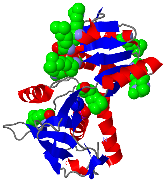
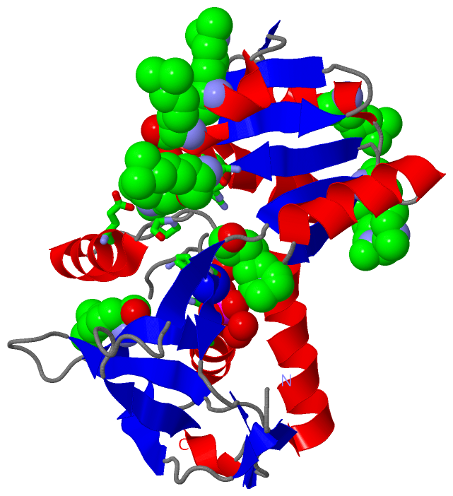
Asymmetric/Biological UnitChain A from PDB Type:PROTEIN Length:232 aligned with Q8U3S5_PYRFU | Q8U3S5 from UniProtKB/TrEMBL Length:242 Alignment length:232 20 30 40 50 60 70 80 90 100 110 120 130 140 150 160 170 180 190 200 210 220 230 240 Q8U3S5_PYRFU 11 IPVKKVEYVFIELDKMKPHEQLVQRELEDFIESVTGSGIFWKPMLLAKIPGTDEYLIVDGHHRWAGLQKLGAKRAPSVILDYFDEGVKVYTWYPAFKGDVNKVIERLKAEGLEVIEDEKAEEKAEKGEIAFALIGEKSFAIPGGLEEQKKVSKVLDEMDQAKEIELVYYGLKEDAKADMEKGEIDYVFIRKAPTKEEVMELVKRGEVFSPKTTRHVLPFIPDKIDVKLEDLF 242 SCOP domains d1vk1a_ A: Hypothetical protein PF0380 SCOP domains CATH domains 1vk1A01 A:11-96,A:227-242 1vk1A02 A:97-226 Conserved hypothetical protein from pyrococcus furiosus pfu- 392566-001, domain 2 1vk1A01 CATH domains Pfam domains --------ParBc-1vk1A01 A:19-114 -------------------------------------------------------------------------------------------------------------------------------- Pfam domains SAPs(SNPs) ---------------------------------------------------------------------------------------------------------------------------------------------------------------------------------------------------------------------------------------- SAPs(SNPs) PROSITE ---------------------------------------------------------------------------------------------------------------------------------------------------------------------------------------------------------------------------------------- PROSITE Transcript ---------------------------------------------------------------------------------------------------------------------------------------------------------------------------------------------------------------------------------------- Transcript 1vk1 A 11 IPVKKVEYVFIELDKMkPHEQLVQRELEDFIESVTGSGIFWKPMLLAKIPGTDEYLIVDGHHRWAGLQKLGAKRAPSVILDYFDEGVKVYTWYPAFkGDVNkVIERLKAEGLEVIEDEKAEEkAEkGEIAFALIGEKSFAIPGGLEEQkKVSKVLDEMDQAkEIELVYYGLKEDAKADMEKGEIDYVFIRkAPTKEEVMELVKRGEVFSPkTTRHVLPFIPDKIDVKLEDLF 242 20 | 30 40 50 60 70 80 90 100 |110 | 120 130 | | 140 150 160 170 | 180 190 200| 210 220| 230 240 27-MLY 107-MLY| 133-MLY 159-MLY 172-MLY 201-MLY 221-MLY 112-MLY 136-MLY
|
||||||||||||||||||||
SCOP Domains (1, 1)
Asymmetric/Biological Unit
 |
CATH Domains (2, 2)
Asymmetric/Biological Unit
|
Pfam Domains (1, 1)
Asymmetric/Biological Unit
|
Gene Ontology (0, 0)|
Asymmetric/Biological Unit(hide GO term definitions)
(no "Gene Ontology" information available for 1VK1)
|
Interactive Views
|
|||||||||||||||||||||||||||||||||||||||||||||||||||||||||||||||||||||||||||||||||||||||||||||||||||||||||||||||||||||||||||||||||||||||||||||||||||||||||
Still Images
|
||||||||||||||||
Databases
|
||||||||||||||||||||||||||||||||||||||||||||||||||||||||||||||||||||||||||||||||||||||||||||||||||||||||||||||||||||||||||||||||||||||||||||||||||||||||||||||||
Analysis Tools
|
|||||||||||||||||||||||||||||||||||||||||||||||||||||||||||||
Entries Sharing at Least One Protein Chain (UniProt ID)
Related Entries Specified in the PDB File
|
|