| molecular function |
|---|
| | GO:0042803 | | protein homodimerization activity | | Interacting selectively and non-covalently with an identical protein to form a homodimer. |
| biological process |
|---|
| | GO:0007409 | | axonogenesis | | De novo generation of a long process of a neuron, that carries efferent (outgoing) action potentials from the cell body towards target cells. Refers to the morphogenesis or creation of shape or form of the developing axon. |
| | GO:0007420 | | brain development | | The process whose specific outcome is the progression of the brain over time, from its formation to the mature structure. Brain development begins with patterning events in the neural tube and ends with the mature structure that is the center of thought and emotion. The brain is responsible for the coordination and control of bodily activities and the interpretation of information from the senses (sight, hearing, smell, etc.). |
| | GO:0007155 | | cell adhesion | | The attachment of a cell, either to another cell or to an underlying substrate such as the extracellular matrix, via cell adhesion molecules. |
| | GO:0001709 | | cell fate determination | | A process involved in cell fate commitment. Once determination has taken place, a cell becomes committed to differentiate down a particular pathway regardless of its environment. |
| | GO:0007417 | | central nervous system development | | The process whose specific outcome is the progression of the central nervous system over time, from its formation to the mature structure. The central nervous system is the core nervous system that serves an integrating and coordinating function. In vertebrates it consists of the brain and spinal cord. In those invertebrates with a central nervous system it typically consists of a brain, cerebral ganglia and a nerve cord. |
| | GO:0009953 | | dorsal/ventral pattern formation | | The regionalization process in which the areas along the dorsal/ventral axis are established that will lead to differences in cell differentiation. The dorsal/ventral axis is defined by a line that runs orthogonal to both the anterior/posterior and left/right axes. The dorsal end is defined by the upper or back side of an organism. The ventral end is defined by the lower or front side of an organism. |
| | GO:0048704 | | embryonic skeletal system morphogenesis | | The process in which the anatomical structures of the skeleton are generated and organized during the embryonic phase. |
| | GO:0007156 | | homophilic cell adhesion via plasma membrane adhesion molecules | | The attachment of a plasma membrane adhesion molecule in one cell to an identical molecule in an adjacent cell. |
| | GO:0007399 | | nervous system development | | The process whose specific outcome is the progression of nervous tissue over time, from its formation to its mature state. |
| cellular component |
|---|
| | GO:0030054 | | cell junction | | A cellular component that forms a specialized region of connection between two or more cells or between a cell and the extracellular matrix. At a cell junction, anchoring proteins extend through the plasma membrane to link cytoskeletal proteins in one cell to cytoskeletal proteins in neighboring cells or to proteins in the extracellular matrix. |
| | GO:0009986 | | cell surface | | The external part of the cell wall and/or plasma membrane. |
| | GO:0005615 | | extracellular space | | That part of a multicellular organism outside the cells proper, usually taken to be outside the plasma membranes, and occupied by fluid. |
| | GO:0016021 | | integral component of membrane | | The component of a membrane consisting of the gene products and protein complexes having at least some part of their peptide sequence embedded in the hydrophobic region of the membrane. |
| | GO:0016020 | | membrane | | A lipid bilayer along with all the proteins and protein complexes embedded in it an attached to it. |
| | GO:0005886 | | plasma membrane | | The membrane surrounding a cell that separates the cell from its external environment. It consists of a phospholipid bilayer and associated proteins. |
| | GO:0045202 | | synapse | | The junction between a nerve fiber of one neuron and another neuron, muscle fiber or glial cell. As the nerve fiber approaches the synapse it enlarges into a specialized structure, the presynaptic nerve ending, which contains mitochondria and synaptic vesicles. At the tip of the nerve ending is the presynaptic membrane; facing it, and separated from it by a minute cleft (the synaptic cleft) is a specialized area of membrane on the receiving cell, known as the postsynaptic membrane. In response to the arrival of nerve impulses, the presynaptic nerve ending secretes molecules of neurotransmitters into the synaptic cleft. These diffuse across the cleft and transmit the signal to the postsynaptic membrane. |
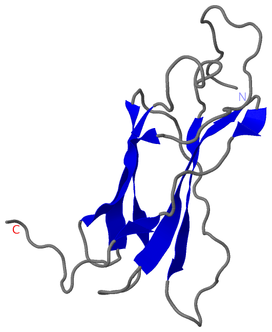
 Description
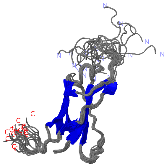