| molecular function |
|---|
| | GO:0008269 | | JAK pathway signal transduction adaptor activity | | The binding activity of a molecule that brings together two molecules of the JAK signal transduction pathway, permitting them to function in a coordinated way. |
| | GO:0005070 | | SH3/SH2 adaptor activity | | Interacting selectively and non-covalently and simultaneously with one or more signal transduction molecules, usually acting as a scaffold to bring these molecules into close proximity either using their own SH2/SH3 domains (e.g. Grb2) or those of their target molecules (e.g. SAM68). |
| | GO:0042802 | | identical protein binding | | Interacting selectively and non-covalently with an identical protein or proteins. |
| | GO:0005515 | | protein binding | | Interacting selectively and non-covalently with any protein or protein complex (a complex of two or more proteins that may include other nonprotein molecules). |
| | GO:0004871 | | signal transducer activity | | Conveys a signal across a cell to trigger a change in cell function or state. A signal is a physical entity or change in state that is used to transfer information in order to trigger a response. |
| | GO:0035591 | | signaling adaptor activity | | The binding activity of a molecule that brings together two or more molecules in a signaling pathway, permitting those molecules to function in a coordinated way. Adaptor molecules themselves do not have catalytic activity. |
| | GO:0005068 | | transmembrane receptor protein tyrosine kinase adaptor activity | | The binding activity of a molecule that brings together a transmembrane receptor protein tyrosine kinase and one or more other molecules, permitting them to function in a coordinated way. |
| biological process |
|---|
| | GO:0001922 | | B-1 B cell homeostasis | | The process of regulating the proliferation and elimination of B cells of the B-1 subset such that the total number of B-1 B cells within a whole or part of an organism is stable over time in the absence of an outside stimulus. B-1 B cells are a distinct subset of B cells characterized as being CD5 positive, found predominantly in the peritoneum, pleural cavities, and spleen, and enriched for self-reactivity. |
| | GO:0030036 | | actin cytoskeleton organization | | A process that is carried out at the cellular level which results in the assembly, arrangement of constituent parts, or disassembly of cytoskeletal structures comprising actin filaments and their associated proteins. |
| | GO:0050851 | | antigen receptor-mediated signaling pathway | | A series of molecular signals initiated by the cross-linking of an antigen receptor on a B or T cell. |
| | GO:0050873 | | brown fat cell differentiation | | The process in which a relatively unspecialized cell acquires specialized features of a brown adipocyte, an animal connective tissue cell involved in adaptive thermogenesis. Brown adipocytes contain multiple small droplets of triglycerides and a high number of mitochondria. |
| | GO:0019221 | | cytokine-mediated signaling pathway | | A series of molecular signals initiated by the binding of a cytokine to a receptor on the surface of a cell, and ending with regulation of a downstream cellular process, e.g. transcription. |
| | GO:0042593 | | glucose homeostasis | | Any process involved in the maintenance of an internal steady state of glucose within an organism or cell. |
| | GO:0008286 | | insulin receptor signaling pathway | | The series of molecular signals generated as a consequence of the insulin receptor binding to insulin. |
| | GO:0035556 | | intracellular signal transduction | | The process in which a signal is passed on to downstream components within the cell, which become activated themselves to further propagate the signal and finally trigger a change in the function or state of the cell. |
| | GO:0046325 | | negative regulation of glucose import | | Any process that stops, prevents, or reduces the frequency, rate or extent of the import of the hexose monosaccharide glucose into a cell or organelle. |
| | GO:0007399 | | nervous system development | | The process whose specific outcome is the progression of nervous tissue over time, from its formation to its mature state. |
| | GO:0009967 | | positive regulation of signal transduction | | Any process that activates or increases the frequency, rate or extent of signal transduction. |
| | GO:0046425 | | regulation of JAK-STAT cascade | | Any process that modulates the frequency, rate or extent of the JAK-STAT signaling pathway. |
| | GO:0046578 | | regulation of Ras protein signal transduction | | Any process that modulates the frequency, rate or extent of Ras protein signal transduction. |
| | GO:0050776 | | regulation of immune response | | Any process that modulates the frequency, rate or extent of the immune response, the immunological reaction of an organism to an immunogenic stimulus. |
| | GO:0019222 | | regulation of metabolic process | | Any process that modulates the frequency, rate or extent of the chemical reactions and pathways within a cell or an organism. |
| | GO:0007165 | | signal transduction | | The cellular process in which a signal is conveyed to trigger a change in the activity or state of a cell. Signal transduction begins with reception of a signal (e.g. a ligand binding to a receptor or receptor activation by a stimulus such as light), or for signal transduction in the absence of ligand, signal-withdrawal or the activity of a constitutively active receptor. Signal transduction ends with regulation of a downstream cellular process, e.g. regulation of transcription or regulation of a metabolic process. Signal transduction covers signaling from receptors located on the surface of the cell and signaling via molecules located within the cell. For signaling between cells, signal transduction is restricted to events at and within the receiving cell. |
| cellular component |
|---|
| | GO:0005884 | | actin filament | | A filamentous structure formed of a two-stranded helical polymer of the protein actin and associated proteins. Actin filaments are a major component of the contractile apparatus of skeletal muscle and the microfilaments of the cytoskeleton of eukaryotic cells. The filaments, comprising polymerized globular actin molecules, appear as flexible structures with a diameter of 5-9 nm. They are organized into a variety of linear bundles, two-dimensional networks, and three dimensional gels. In the cytoskeleton they are most highly concentrated in the cortex of the cell just beneath the plasma membrane. |
| | GO:0005737 | | cytoplasm | | All of the contents of a cell excluding the plasma membrane and nucleus, but including other subcellular structures. |
| | GO:0016020 | | membrane | | A lipid bilayer along with all the proteins and protein complexes embedded in it an attached to it. |
| | GO:0005886 | | plasma membrane | | The membrane surrounding a cell that separates the cell from its external environment. It consists of a phospholipid bilayer and associated proteins. |
| | GO:0001726 | | ruffle | | Projection at the leading edge of a crawling cell; the protrusions are supported by a microfilament meshwork. |
| | GO:0001725 | | stress fiber | | A contractile actin filament bundle that consists of short actin filaments with alternating polarity, cross-linked by alpha-actinin and possibly other actin bundling proteins, and with myosin present in a periodic distribution along the fiber. |
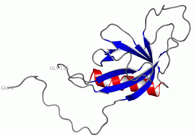
 Description
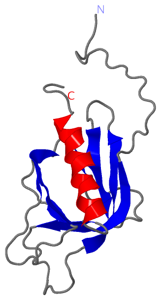
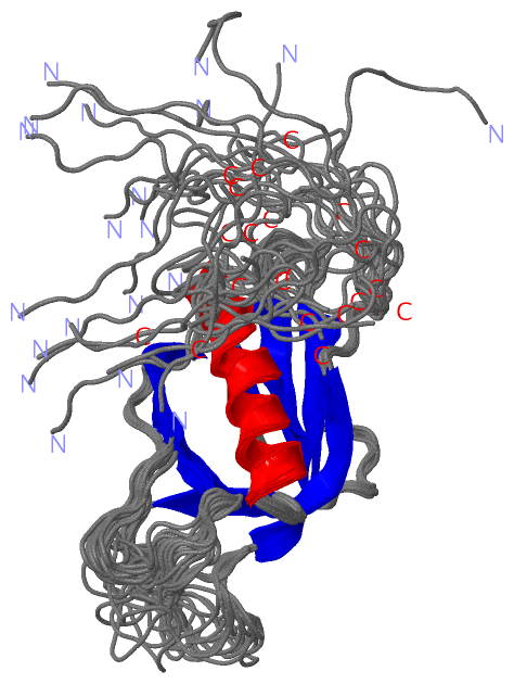