|
|
|
|
 Description
|
|
Compounds
|
||||||||||||||||||||||||||||||||||||||||||||||||||
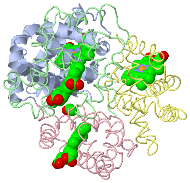
Chains, Units
Summary Information (see also Sequences/Alignments below) |
Ligands, Modified Residues, Ions (2, 6)| Asymmetric/Biological Unit (2, 6) |
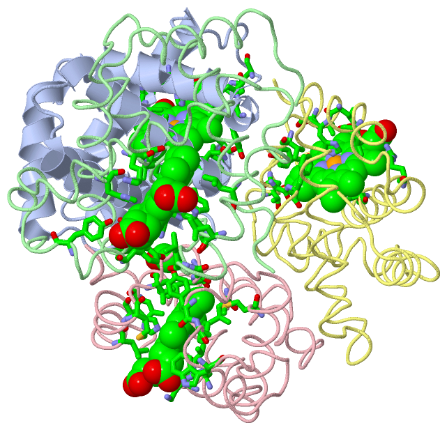
Sites (4, 4)
Asymmetric Unit (4, 4)
|
SS Bonds (0, 0)| (no "SS Bond" information available for 1V4X) |
Cis Peptide Bonds (0, 0)| (no "Cis Peptide Bond" information available for 1V4X) |
SAPs(SNPs)/Variants (0, 0)| (no "SAP(SNP)/Variant" information available for 1V4X) |
PROSITE Motifs (0, 0)| (no "PROSITE Motif" information available for 1V4X) |
Exons (0, 0)| (no "Exon" information available for 1V4X) |
Sequences/Alignments
Asymmetric/Biological UnitChain A from PDB Type:PROTEIN Length:144 aligned with Q8AYM0_THUTH | Q8AYM0 from UniProtKB/TrEMBL Length:144 Alignment length:144 10 20 30 40 50 60 70 80 90 100 110 120 130 140 Q8AYM0_THUTH 1 MTTLSDKDKSTVKALWGKISKSADAIGADALGRMLAVYPQTKTYFSHWPDMSPGSGPVKAHGKKVMGGVALAVSKIDDLTTGLGDLSELHAFKMRVDPSNFKILSHCILVVVAKMFPKEFTPDAHVSLDKFLASVALALAERYR 144 SCOP domains d1v4xa_ A: Hemoglobin, alpha-chain SCOP domains CATH domains -1v4xA00 A:1-143 Globins CATH domains Pfam domains ------------------------------------------------------------------------------------------------------------------------------------------------ Pfam domains SAPs(SNPs) ------------------------------------------------------------------------------------------------------------------------------------------------ SAPs(SNPs) PROSITE ------------------------------------------------------------------------------------------------------------------------------------------------ PROSITE Transcript ------------------------------------------------------------------------------------------------------------------------------------------------ Transcript 1v4x A 0 xTTLSDKDKSTVKALWGKISKSADAIGADALGRMLAVYPQTKTYFSHWPDMSPGSGPVKAHGKKVMGGVALAVSKIDDLTTGLGDLSELHAFKMRVDPSNFKILSHCILVVVAKMFPKEFTPDAHVSLDKFLASVALALAERYR 143 | 9 19 29 39 49 59 69 79 89 99 109 119 129 139 | 0-ACE Chain B from PDB Type:PROTEIN Length:146 aligned with Q8AYM1_THUTH | Q8AYM1 from UniProtKB/TrEMBL Length:147 Alignment length:146 11 21 31 41 51 61 71 81 91 101 111 121 131 141 Q8AYM1_THUTH 2 VEWTQQERSIIAGIFANLNYEDIGPKALARCLIVYPWTQRYFGAYGDLSTPDAIKGNAKIAAHGVKVLHGLDRAVKNMDNINEAYSELSVLHSDKLHVDPDNFRILGDCLTVVIAANLGDAFTVETQCAFQKFLAVVVFALGRKYH 147 SCOP domains d1v4xb_ B: Hemoglobin, beta-chain SCOP domains CATH domains 1v4xB00 B:201-346 Globins CATH domains Pfam domains -------------------------------------------------------------------------------------------------------------------------------------------------- Pfam domains SAPs(SNPs) -------------------------------------------------------------------------------------------------------------------------------------------------- SAPs(SNPs) PROSITE -------------------------------------------------------------------------------------------------------------------------------------------------- PROSITE Transcript -------------------------------------------------------------------------------------------------------------------------------------------------- Transcript 1v4x B 201 VEWTQQERSIIAGIFANLNYEDIGPKALARCLIVYPWTQRYFGAYGDLSTPDAIKGNAKIAAHGVKVLHGLDRAVKNMDNINEAYSELSVLHSDKLHVDPDNFRILGDCLTVVIAANLGDAFTVETQCAFQKFLAVVVFALGRKYH 346 210 220 230 240 250 260 270 280 290 300 310 320 330 340 Chain C from PDB Type:PROTEIN Length:144 aligned with Q8AYM0_THUTH | Q8AYM0 from UniProtKB/TrEMBL Length:144 Alignment length:144 10 20 30 40 50 60 70 80 90 100 110 120 130 140 Q8AYM0_THUTH 1 MTTLSDKDKSTVKALWGKISKSADAIGADALGRMLAVYPQTKTYFSHWPDMSPGSGPVKAHGKKVMGGVALAVSKIDDLTTGLGDLSELHAFKMRVDPSNFKILSHCILVVVAKMFPKEFTPDAHVSLDKFLASVALALAERYR 144 SCOP domains d1v4xc_ C: Hemoglobin, alpha-chain SCOP domains CATH domains -1v4xC00 C:401-543 Globins CATH domains Pfam domains ------------------------------------------------------------------------------------------------------------------------------------------------ Pfam domains SAPs(SNPs) ------------------------------------------------------------------------------------------------------------------------------------------------ SAPs(SNPs) PROSITE ------------------------------------------------------------------------------------------------------------------------------------------------ PROSITE Transcript ------------------------------------------------------------------------------------------------------------------------------------------------ Transcript 1v4x C 400 xTTLSDKDKSTVKALWGKISKSADAIGADALGRMLAVYPQTKTYFSHWPDMSPGSGPVKAHGKKVMGGVALAVSKIDDLTTGLGDLSELHAFKMRVDPSNFKILSHCILVVVAKMFPKEFTPDAHVSLDKFLASVALALAERYR 543 | 409 419 429 439 449 459 469 479 489 499 509 519 529 539 400-ACE Chain D from PDB Type:PROTEIN Length:146 aligned with Q8AYM1_THUTH | Q8AYM1 from UniProtKB/TrEMBL Length:147 Alignment length:146 11 21 31 41 51 61 71 81 91 101 111 121 131 141 Q8AYM1_THUTH 2 VEWTQQERSIIAGIFANLNYEDIGPKALARCLIVYPWTQRYFGAYGDLSTPDAIKGNAKIAAHGVKVLHGLDRAVKNMDNINEAYSELSVLHSDKLHVDPDNFRILGDCLTVVIAANLGDAFTVETQCAFQKFLAVVVFALGRKYH 147 SCOP domains d1v4xd_ D: Hemoglobin, beta-chain SCOP domains CATH domains 1v4xD00 D:601-746 Globins CATH domains Pfam domains -------------------------------------------------------------------------------------------------------------------------------------------------- Pfam domains SAPs(SNPs) -------------------------------------------------------------------------------------------------------------------------------------------------- SAPs(SNPs) PROSITE -------------------------------------------------------------------------------------------------------------------------------------------------- PROSITE Transcript -------------------------------------------------------------------------------------------------------------------------------------------------- Transcript 1v4x D 601 VEWTQQERSIIAGIFANLNYEDIGPKALARCLIVYPWTQRYFGAYGDLSTPDAIKGNAKIAAHGVKVLHGLDRAVKNMDNINEAYSELSVLHSDKLHVDPDNFRILGDCLTVVIAANLGDAFTVETQCAFQKFLAVVVFALGRKYH 746 610 620 630 640 650 660 670 680 690 700 710 720 730 740
|
||||||||||||||||||||
SCOP Domains (2, 4)
Asymmetric/Biological Unit
 |
CATH Domains (1, 4)
Asymmetric/Biological Unit
|
Pfam Domains (0, 0)| (no "Pfam Domain" information available for 1V4X) |
Gene Ontology (8, 16)|
Asymmetric/Biological Unit(hide GO term definitions) Chain A,C (Q8AYM0_THUTH | Q8AYM0)
Chain B,D (Q8AYM1_THUTH | Q8AYM1)
|
||||||||||||||||||||||||||||||||||||||||||||||||||||||||||||||||||||||||||||||||||||||||||||||||||||||||||||||||||||||||||||||||||||
Interactive Views
|
||||||||||||||||||||||||||||||||||||||||||||||||||||||||||||||||||||||||||||||||||||||||||||||||||||||||||||||||||||||||||||||||||||||||||||||||||
Still Images
|
||||||||||||||||
Databases
|
||||||||||||||||||||||||||||||||||||||||||||||||||||||||||||||||||||||||||||||||||||||||||||||||||||||||||||||||||||||||||||||||||||||||||||||||||||||||||||||||||||||||||||||||||||||||||
Analysis Tools
|
||||||||||||||||||||||||||||||||||||||||||||||||||||||||||||||||||||||||
Entries Sharing at Least One Protein Chain (UniProt ID)
Related Entries Specified in the PDB File
|
|