Show PDB file:   
         Plain Text   HTML   (compressed file size)
QuickSearch:   
by PDB,NDB,UniProt,PROSITE Code or Search Term(s)  
(-)Asymmetric Unit
(-)Biological Unit 1
(-)Biological Unit 2
collapse expand < >
Image Asymmetric Unit
Asymmetric Unit  (Jmol Viewer)
Image Biological Unit 1
Biological Unit 1  (Jmol Viewer)
Image Biological Unit 2
Biological Unit 2  (Jmol Viewer)

(-) Description

Title :  THE CRYSTAL STRUCTURE OF PEPTIDE DEFORMYLASE FROM THERMUS THERMOPHILUS HB8
 
Authors :  M. Kamo, N. Kudo, W. C. Lee, K. Ito, H. Motoshim, M. Tanokura, Riken Structural Genomics/Proteomics Initiative (Rsgi)
Date :  07 Nov 03  (Deposition) - 28 Dec 04  (Release) - 24 Feb 09  (Revision)
Method :  X-RAY DIFFRACTION
Resolution :  1.81
Chains :  Asym. Unit :  A,B
Biol. Unit 1:  A  (1x)
Biol. Unit 2:  B  (1x)
Keywords :  Peptide Deformylase, Protein Synthesis, Riken Structural Genomics/Proteomics Initiative, Rsgi, Structural Genomics, Hydrolase (Keyword Search: [Gene Ontology, PubMed, Web (Google))
 
Reference :  M. Kamo, N. Kudo, W. C. Lee, K. Ito, H. Motoshim, M. Tanokura
The Crystal Structure Of Peptide Deformylase From Thermus Thermophilus Hb8
To Be Published
PubMed: search
(for further references see the PDB file header)

(-) Compounds

Molecule 1 - PEPTIDE DEFORMYLASE
    ChainsA, B
    EC Number3.5.1.88
    EngineeredYES
    Expression SystemESCHERICHIA COLI
    Expression System PlasmidPET11A
    Expression System StrainBL21(DE3) PLYS S
    Expression System Taxid562
    Expression System Vector TypePLASMID
    Organism ScientificTHERMUS THERMOPHILUS
    Organism Taxid274
    SynonymPDF, POLYPEPTIDE DEFORMYLASE

 Structural Features

(-) Chains, Units

  12
Asymmetric Unit AB
Biological Unit 1 (1x)A 
Biological Unit 2 (1x) B

Summary Information (see also Sequences/Alignments below)

(-) Ligands, Modified Residues, Ions  (1, 2)

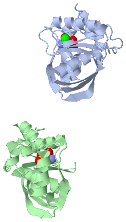
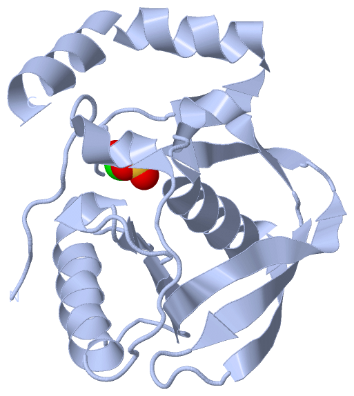
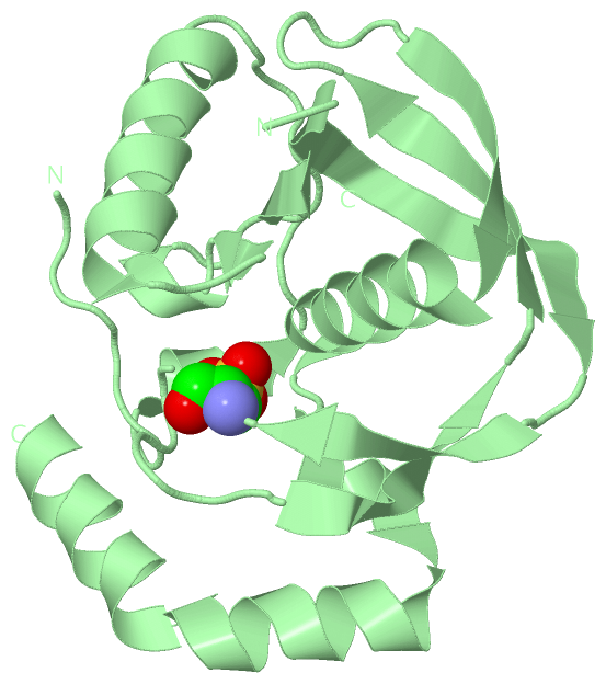
Asymmetric Unit (1, 2)
No.NameCountTypeFull Name
1OCS2Mod. Amino AcidCYSTEINESULFONIC ACID
Biological Unit 1 (1, 1)
No.NameCountTypeFull Name
1OCS1Mod. Amino AcidCYSTEINESULFONIC ACID
Biological Unit 2 (1, 1)
No.NameCountTypeFull Name
1OCS1Mod. Amino AcidCYSTEINESULFONIC ACID

(-) Sites  (0, 0)

(no "Site" information available for 1V3Y)

(-) SS Bonds  (0, 0)

(no "SS Bond" information available for 1V3Y)

(-) Cis Peptide Bonds  (0, 0)

(no "Cis Peptide Bond" information available for 1V3Y)

 Sequence-Structure Mapping

(-) SAPs(SNPs)/Variants  (0, 0)

(no "SAP(SNP)/Variant" information available for 1V3Y)

(-) PROSITE Motifs  (0, 0)

(no "PROSITE Motif" information available for 1V3Y)

(-) Exons   (0, 0)

(no "Exon" information available for 1V3Y)

(-) Sequences/Alignments

Asymmetric Unit
   Reformat: Number of residues per line =  ('0' or empty: single-line sequence representation)
  Number of residues per labelling interval =   
  UniProt sequence: complete  aligned part    
   Show mapping: SCOP domains CATH domains Pfam domains Secondary structure (by author)
SAPs(SNPs) PROSITE motifs Exons
(details for a mapped element are shown in a popup box when the mouse pointer rests over it)
Chain A from PDB  Type:PROTEIN  Length:170
 aligned with DEF_THETH | P43522 from UniProtKB/Swiss-Prot  Length:192

    Alignment length:182
                                    10        20        30        40        50        60        70        80        90       100       110       120       130       140       150       160       170       180  
            DEF_THETH     1 MVYPIRLYGDPVLRRKARPVEDFSGIKRLAEDMLETMFEAKGVGLAAPQIGLSQRLFVAVEYADEPEGEEERPLRELVRRVYVVANPVITYREGLVEGTEGCLSLPGLYSEEVPRAERIRVEYQDEEGRGRVLELEGYMARVFQHEIDHLDGILFFERLPKPKREAFLEANRAELVRFQKEA 182
               SCOP domains d1v3ya_ A: Peptide deformylase                                                                                                                                                         SCOP domains
               CATH domains 1v3yA00 A:1-182 Peptide Deformylase                                                                                                                                                    CATH domains
               Pfam domains -------------------------------------------------------------------------------------------------------------------------------------------------------------------------------------- Pfam domains
         Sec.struct. author .........hhhhhh.........hhhhhhhhhhhhhhhh...eee.hhhh....eeeee.------------.......eeeeeeeeeeeee..eeeee.........eeeeee..eeeeeee.....eeeeeeehhhhhhhhhhhhhhh..hhhhhhhhhhhhhhhhhhhhhhhhhhhhh Sec.struct. author
                 SAPs(SNPs) -------------------------------------------------------------------------------------------------------------------------------------------------------------------------------------- SAPs(SNPs)
                    PROSITE -------------------------------------------------------------------------------------------------------------------------------------------------------------------------------------- PROSITE
                 Transcript -------------------------------------------------------------------------------------------------------------------------------------------------------------------------------------- Transcript
                 1v3y A   1 MVYPIRLYGDPVLRRKARPVEDFSGIKRLAEDMLETMFEAKGVGLAAPQIGLSQRLFVAVE------------LRELVRRVYVVANPVITYREGLVEGTEGcLSLPGLYSEEVPRAERIRVEYQDEEGRGRVLELEGYMARVFQHEIDHLDGILFFERLPKPKREAFLEANRAELVRFQKEA 182
                                    10        20        30        40        50        60|        -   |    80        90       100 |     110       120       130       140       150       160       170       180  
                                                                                       61           74                         102-OCS                                                                            

Chain B from PDB  Type:PROTEIN  Length:167
 aligned with DEF_THETH | P43522 from UniProtKB/Swiss-Prot  Length:192

    Alignment length:182
                                    11        21        31        41        51        61        71        81        91       101       111       121       131       141       151       161       171       181  
            DEF_THETH     2 VYPIRLYGDPVLRRKARPVEDFSGIKRLAEDMLETMFEAKGVGLAAPQIGLSQRLFVAVEYADEPEGEEERPLRELVRRVYVVANPVITYREGLVEGTEGCLSLPGLYSEEVPRAERIRVEYQDEEGRGRVLELEGYMARVFQHEIDHLDGILFFERLPKPKREAFLEANRAELVRFQKEAR 183
               SCOP domains d1v3yb_ B: Peptide deformylase                                                                                                                                                         SCOP domains
               CATH domains 1v3yB00 B:2-183 Peptide Deformylase                                                                                                                                                    CATH domains
           Pfam domains (1) Pep_deformylase-1v3yB01 B:2-165                                                                                                                                     ------------------ Pfam domains (1)
           Pfam domains (2) Pep_deformylase-1v3yB02 B:2-165                                                                                                                                     ------------------ Pfam domains (2)
         Sec.struct. author ........hhhhhh.........hhhhhhhhhhhhhhhh...eee.hhhh....eeeeee.---------------..eeeeeeeeeeeeee..eeeee.........eeeeee..eeeeeee.....eeeeeeehhhhhhhhhhhhhhh..hhhhhhhhhhhhhhhhhhhhhhhhhhhhhh Sec.struct. author
                 SAPs(SNPs) -------------------------------------------------------------------------------------------------------------------------------------------------------------------------------------- SAPs(SNPs)
                    PROSITE -------------------------------------------------------------------------------------------------------------------------------------------------------------------------------------- PROSITE
                 Transcript -------------------------------------------------------------------------------------------------------------------------------------------------------------------------------------- Transcript
                 1v3y B   2 VYPIRLYGDPVLRRKARPVEDFSGIKRLAEDMLETMFEAKGVGLAAPQIGLSQRLFVAVEY---------------VRRVYVVANPVITYREGLVEGTEGcLSLPGLYSEEVPRAERIRVEYQDEEGRGRVLELEGYMARVFQHEIDHLDGILFFERLPKPKREAFLEANRAELVRFQKEAR 183
                                    11        21        31        41        51        61|        -      | 81        91       101|      111       121       131       141       151       161       171       181  
                                                                                       62              78                     102-OCS                                                                             

   Legend:   → Mismatch (orange background)
  - → Gap (green background, '-', border residues have a numbering label)
    → Modified Residue (blue background, lower-case, 'x' indicates undefined single-letter code, labelled with number + name)
  x → Chemical Group (purple background, 'x', labelled with number + name, e.g. ACE or NH2)
  extra numbering lines below/above indicate numbering irregularities and modified residue names etc., number ends below/above '|'

 Classification and Annotation

(-) SCOP Domains  (1, 2)

Asymmetric Unit

(-) CATH Domains  (1, 2)

Asymmetric Unit
(-)
Class: Alpha Beta (26913)

(-) Pfam Domains  (1, 2)

Asymmetric Unit

(-) Gene Ontology  (5, 5)

Asymmetric Unit(hide GO term definitions)
Chain A,B   (DEF_THETH | P43522)
molecular function
    GO:0016787    hydrolase activity    Catalysis of the hydrolysis of various bonds, e.g. C-O, C-N, C-C, phosphoric anhydride bonds, etc. Hydrolase is the systematic name for any enzyme of EC class 3.
    GO:0005506    iron ion binding    Interacting selectively and non-covalently with iron (Fe) ions.
    GO:0046872    metal ion binding    Interacting selectively and non-covalently with any metal ion.
    GO:0042586    peptide deformylase activity    Catalysis of the reaction: formyl-L-methionyl peptide + H2O = formate + methionyl peptide.
biological process
    GO:0006412    translation    The cellular metabolic process in which a protein is formed, using the sequence of a mature mRNA or circRNA molecule to specify the sequence of amino acids in a polypeptide chain. Translation is mediated by the ribosome, and begins with the formation of a ternary complex between aminoacylated initiator methionine tRNA, GTP, and initiation factor 2, which subsequently associates with the small subunit of the ribosome and an mRNA or circRNA. Translation ends with the release of a polypeptide chain from the ribosome.

 Visualization

(-) Interactive Views

Asymmetric Unit
  Complete Structure
    Jena3D(integrated viewing of ligand, site, SAP, PROSITE, SCOP information)
    WebMol | AstexViewer[tm]@PDBe
(Java Applets, require no local installation except for Java; loading may be slow)
    STRAP
(Java WebStart application, automatic local installation, requires Java; full application with system access!)
    RasMol
(require local installation)
    Molscript (VRML)
(requires installation of a VRML viewer; select preferred view via VRML and generate a mono or stereo PDF format file)
 
  Ligands, Modified Residues, Ions
    OCS  [ RasMol | Jena3D ]  +environment [ RasMol | Jena3D ]
 
  Sites
(no "Sites" information available for 1v3y)
 
  Cis Peptide Bonds
(no "Cis Peptide Bonds" information available for 1v3y)
 
Biological Units
  Complete Structure
    Biological Unit 1  [ Jena3D ]
    Biological Unit 2  [ Jena3D ]

(-) Still Images

Jmol
  protein: cartoon or spacefill or dots and stick; nucleic acid: cartoon and stick; ligands: spacefill; active site: stick
Molscript
  protein, nucleic acid: cartoon; ligands: spacefill; active site: ball and stick

 Databases and Analysis Tools

(-) Databases

Access by PDB/NDB ID
  1v3y
    Family and Domain InformationProDom | SYSTERS
    General Structural InformationGlycoscienceDB | MMDB | NDB | OCA | PDB | PDBe | PDBj | PDBsum | PDBWiki | PQS | PROTEOPEDIA
    Orientation in MembranesOPM
    Protein SurfaceSURFACE
    Secondary StructureDSSP (structure derived) | HSSP (homology derived)
    Structural GenomicsGeneCensus
    Structural NeighboursCE | VAST
    Structure ClassificationCATH | Dali | SCOP
    Validation and Original DataBMRB Data View | BMRB Restraints Grid | EDS | PROCHECK | RECOORD | WHAT_CHECK
 
Access by UniProt ID/Accession number
  DEF_THETH | P43522
    Comparative Protein Structure ModelsModBase
    Genomic InformationEnsembl
    Protein-protein InteractionDIP
    Sequence, Family and Domain InformationInterPro | Pfam | SMART | UniProtKB/SwissProt
 
Access by Enzyme Classificator   (EC Number)
  3.5.1.88
    General Enzyme InformationBRENDA | EC-PDB | Enzyme | IntEnz
    PathwayKEGG | MetaCyc
 
Access by Disease Identifier   (MIM ID)
  (no 'MIM ID' available)
    Disease InformationOMIM
 
Access by GenAge ID
  (no 'GenAge ID' available)
    Age Related InformationGenAge

(-) Analysis Tools

Access by PDB/NDB ID
    Domain InformationXDom
    Interatomic Contacts of Structural UnitsCSU
    Ligand-protein ContactsLPC
    Protein CavitiescastP
    Sequence and Secondary StructurePDBCartoon
    Structure AlignmentSTRAP(Java WebStart application, automatic local installation, requires Java; full application with system access!)
    Structure and Sequence BrowserSTING
 
Access by UniProt ID/Accession number
  DEF_THETH | P43522
    Protein Disorder PredictionDisEMBL | FoldIndex | GLOBPLOT (for more information see DisProt)

 Related Entries

(-) Entries Sharing at Least One Protein Chain (UniProt ID)

(no "Entries Sharing at Least One Protein Chain" available for 1V3Y)

(-) Related Entries Specified in the PDB File

(no "Related Entries Specified in the PDB File" available for 1V3Y)