|
|
|
|
 Description
|
|
Compounds
|
||||||||||||||||||||||||||||||||||||||||||||||||||||||||
Chains, Units
Summary Information (see also Sequences/Alignments below) |
Ligands, Modified Residues, Ions (2, 4)| Asymmetric Unit (2, 4) Biological Unit 1 (2, 2) Biological Unit 2 (2, 2) |
Sites (4, 4)
Asymmetric Unit (4, 4)
|
SS Bonds (0, 0)| (no "SS Bond" information available for 1V2B) |
Cis Peptide Bonds (0, 0)| (no "Cis Peptide Bond" information available for 1V2B) |
SAPs(SNPs)/Variants (0, 0)| (no "SAP(SNP)/Variant" information available for 1V2B) |
PROSITE Motifs (0, 0)| (no "PROSITE Motif" information available for 1V2B) |
Exons (0, 0)| (no "Exon" information available for 1V2B) |
Sequences/Alignments
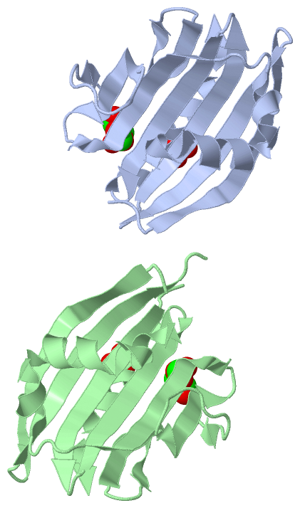
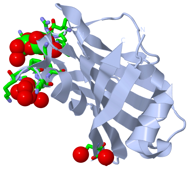
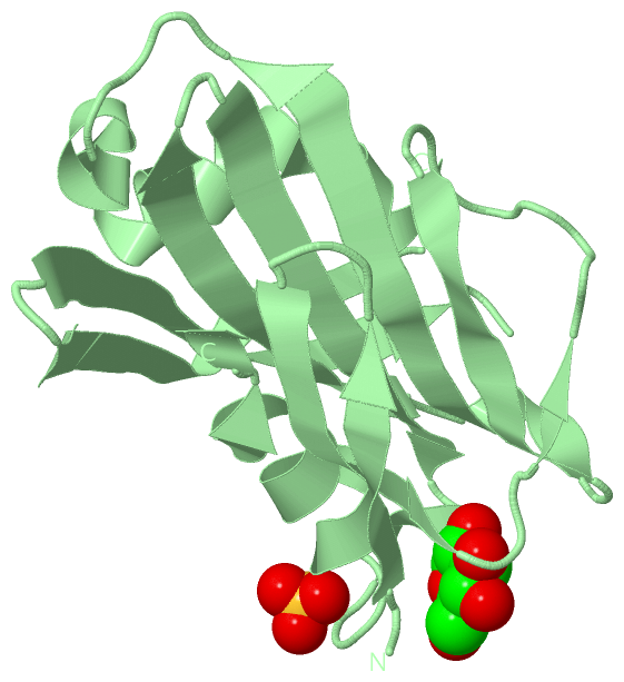
Asymmetric UnitChain A from PDB Type:PROTEIN Length:148 aligned with PSBP2_TOBAC | P18212 from UniProtKB/Swiss-Prot Length:265 Alignment length:171 104 114 124 134 144 154 164 174 184 194 204 214 224 234 244 254 264 PSBP2_TOBAC 95 TDFQTYNGDGFKLQIPSKWNPNKEVEYPGQVLRFEDNFDATSNVIVAITPTDKKSITDFGSPEQFLSQVDYLLGRQAYSGKTDSEGGFESDAVAIANVLETSSAEVGGKPYYYLSVLTRTADGNEGGKHQLITATVNDGKLYICKAQAGDKRWFKGAKKFVENTATSFSLA 265 SCOP domains d1v2ba_ A: Oxygen-evolving enhancer protein PsbP SCOP domains CATH domains 1v2bA00 A:16-186 Protein Transport Mog1p; Chain A CATH domains Pfam domains --------------------------------------------------------------------------------------------------------------------------------------------------------------------------- Pfam domains SAPs(SNPs) --------------------------------------------------------------------------------------------------------------------------------------------------------------------------- SAPs(SNPs) PROSITE --------------------------------------------------------------------------------------------------------------------------------------------------------------------------- PROSITE Transcript --------------------------------------------------------------------------------------------------------------------------------------------------------------------------- Transcript 1v2b A 16 TDFQTYNGDGFKLQIPSKWNPNKEVEYPGQVLRFEDNFDATSNVIVAITPTDKKSITDFGSPEQFLSQVDYLL------------------AVAIANVLETSTAEVGGKQYYYLSILTRT-----GGKHQLVTATVNDGKLYICKAQAGDKRWFKGAKKFVENTATSFSLA 186 25 35 45 55 65 75 85 | - - | 115 125 135 | 145 155 165 175 185 88 107 135 141 Chain B from PDB Type:PROTEIN Length:151 aligned with PSBP2_TOBAC | P18212 from UniProtKB/Swiss-Prot Length:265 Alignment length:171 104 114 124 134 144 154 164 174 184 194 204 214 224 234 244 254 264 PSBP2_TOBAC 95 TDFQTYNGDGFKLQIPSKWNPNKEVEYPGQVLRFEDNFDATSNVIVAITPTDKKSITDFGSPEQFLSQVDYLLGRQAYSGKTDSEGGFESDAVAIANVLETSSAEVGGKPYYYLSVLTRTADGNEGGKHQLITATVNDGKLYICKAQAGDKRWFKGAKKFVENTATSFSLA 265 SCOP domains d1v2bb_ B: Oxygen-evolving enhancer protein PsbP SCOP domains CATH domains 1v2bB00 B:16-186 Protein Transport Mog1p; Chain A CATH domains Pfam domains (1) PsbP-1v2bB01 B:16-186 Pfam domains (1) Pfam domains (2) PsbP-1v2bB02 B:16-186 Pfam domains (2) SAPs(SNPs) --------------------------------------------------------------------------------------------------------------------------------------------------------------------------- SAPs(SNPs) PROSITE --------------------------------------------------------------------------------------------------------------------------------------------------------------------------- PROSITE Transcript --------------------------------------------------------------------------------------------------------------------------------------------------------------------------- Transcript 1v2b B 16 TDFQTYNGDGFKLQIPSKWNPNKEVEYPGQVLRFEDNFDATSNVIVAITPTDKKSITDFGSPEQFLSQVDYLLGR-----------------VAIANVLETSTAEVGGKQYYYLSILTRTAD---GGKHQLVTATVNDGKLYICKAQAGDKRWFKGAKKFVENTATSFSLA 186 25 35 45 55 65 75 85 | - - | 115 125 135 | | 145 155 165 175 185 90 108 137 141
|
||||||||||||||||||||
SCOP Domains (1, 2)
Asymmetric Unit
 |
CATH Domains (1, 2)
Asymmetric Unit
|
Pfam Domains (1, 2)
Asymmetric Unit
|
Gene Ontology (10, 10)|
Asymmetric Unit(hide GO term definitions) Chain A,B (PSBP2_TOBAC | P18212)
|
||||||||||||||||||||||||||||||||||||||||||||||||||||||||||||||||||||||||||||||
Interactive Views
|
|||||||||||||||||||||||||||||||||||||||||||||||||||||||||||||||||||||||||||||||||||||||||||||||||||||||||||||||||||||||||||||||||||||||||||||||||||||||||||||||||||||||||
Still Images
|
||||||||||||||||
Databases
|
||||||||||||||||||||||||||||||||||||||||||||||||||||||||||||||||||||||||||||||||||||||||||||||||||||||||||||||||||||||||||||||||||||||||||||||||||||||||||||||||
Analysis Tools
|
|||||||||||||||||||||||||||||||||||||||||||||||||||||||||||||
Entries Sharing at Least One Protein Chain (UniProt ID)
Related Entries Specified in the PDB File
|
|