Show PDB file:   
         Plain Text   HTML   (compressed file size)
QuickSearch:   
by PDB,NDB,UniProt,PROSITE Code or Search Term(s)  
(-)Asymmetric Unit
(-)Asym. Unit - sites
(-)Biological Unit 1
(-)Biol. Unit 1 - sites
(-)Biological Unit 2
(-)Biological Unit 3
collapse expand < >
Image Asymmetric Unit
Asymmetric Unit  (Jmol Viewer)
Image Asym. Unit - sites
Asym. Unit - sites  (Jmol Viewer)
Image Biological Unit 1
Biological Unit 1  (Jmol Viewer)
Image Biol. Unit 1 - sites
Biol. Unit 1 - sites  (Jmol Viewer)
Image Biological Unit 2
Biological Unit 2  (Jmol Viewer)
Image Biological Unit 3
Biological Unit 3  (Jmol Viewer)

(-) Description

Title :  CRYSTAL STRUCTURE OF D(GCGAAGC), BENDING DUPLEX WITH A BULGE-IN RESIDUE
 
Authors :  T. Sunami, J. Kondo, I. Hirao, K. Watanabe, K. Miura, A. Takenaka
Date :  31 Mar 03  (Deposition) - 09 Mar 04  (Release) - 24 Feb 09  (Revision)
Method :  X-RAY DIFFRACTION
Resolution :  1.60
Chains :  Asym. Unit :  A,B,C,D,E,F
Biol. Unit 1:  A,B  (1x)
Biol. Unit 2:  C,D  (1x)
Biol. Unit 3:  E,F  (1x)
Keywords :  Dna, Sheared G:A Pair, Bulge-In Duplex, G:Axa:G Crossing (Keyword Search: [Gene Ontology, PubMed, Web (Google))
 
Reference :  T. Sunami, J. Kondo, I. Hirao, K. Watanabe, K. Miura, A. Takenaka
Structures Of D(Gcgaagc) And D(Gcgaaagc) (Tetragonal Form): A Switching Of Partners Of The Sheared G. A Pairs To Form A Functional G. Axa. G Crossing.
Acta Crystallogr. , Sect. D V. 60 422 2004
PubMed-ID: 14993665  |  Reference-DOI: 10.1107/S0907444903028415
(for further references see the PDB file header)

(-) Compounds

Molecule 1 - 5'-D(*GP*CP*GP*AP*AP*GP*C)-3'
    ChainsA, B, C, D, E, F
    EngineeredYES
    Other DetailsCHEMICALLY SYNTHESIZED
    SyntheticYES

 Structural Features

(-) Chains, Units

  123456
Asymmetric Unit ABCDEF
Biological Unit 1 (1x)AB    
Biological Unit 2 (1x)  CD  
Biological Unit 3 (1x)    EF

Summary Information (see also Sequences/Alignments below)

(-) Ligands, Modified Residues, Ions  (1, 9)

Asymmetric Unit (1, 9)
No.NameCountTypeFull Name
1NCO9Ligand/IonCOBALT HEXAMMINE(III)
Biological Unit 1 (1, 3)
No.NameCountTypeFull Name
1NCO3Ligand/IonCOBALT HEXAMMINE(III)
Biological Unit 2 (1, 4)
No.NameCountTypeFull Name
1NCO4Ligand/IonCOBALT HEXAMMINE(III)
Biological Unit 3 (1, 2)
No.NameCountTypeFull Name
1NCO2Ligand/IonCOBALT HEXAMMINE(III)

(-) Sites  (9, 9)

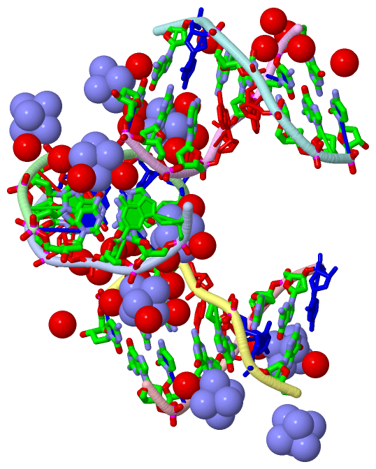
Asymmetric Unit (9, 9)
No.NameEvidenceResiduesDescription
1AC1SOFTWAREDA A:5 , DG A:6 , HOH A:1430 , HOH B:11 , HOH B:37 , DC D:7 , HOH D:10 , HOH D:393BINDING SITE FOR RESIDUE NCO D 400
2AC2SOFTWAREDC A:2 , DG A:3 , HOH A:1405 , HOH A:1433 , HOH A:1443 , HOH A:1456 , DG B:3 , DA D:5BINDING SITE FOR RESIDUE NCO A 1401
3AC3SOFTWAREDG A:3 , DG C:1 , DC C:2 , DA D:5 , DG D:6 , DC D:7 , HOH D:59 , HOH D:65 , HOH D:68 , HOH D:159 , HOH D:209 , HOH D:377BINDING SITE FOR RESIDUE NCO D 402
4AC4SOFTWAREDG B:3 , HOH B:224 , DA E:5 , DG E:6 , DC E:7 , HOH E:39 , HOH E:87 , HOH E:93 , HOH E:94 , DG F:1 , HOH F:110BINDING SITE FOR RESIDUE NCO E 403
5AC5SOFTWAREHOH A:1406 , DA B:5 , DG B:6 , DC B:7 , HOH B:27 , HOH B:30 , HOH B:36 , HOH B:157 , DC E:7 , HOH E:89 , HOH E:129BINDING SITE FOR RESIDUE NCO B 404
6AC6SOFTWAREDG E:1 , HOH E:395 , DA F:5 , DG F:6 , DC F:7 , HOH F:187 , HOH F:268BINDING SITE FOR RESIDUE NCO F 405
7AC7SOFTWAREDA B:5 , HOH B:303 , DC E:2 , DG E:3 , HOH E:234 , DG F:3 , HOH F:394BINDING SITE FOR RESIDUE NCO B 406
8AC8SOFTWAREHOH A:1465 , DA C:5 , DG C:6 , HOH C:174 , DG D:1BINDING SITE FOR RESIDUE NCO C 407
9AC9SOFTWAREDA A:5 , DC C:2 , DG C:3 , HOH C:291 , DG D:3BINDING SITE FOR RESIDUE NCO C 408

(-) SS Bonds  (0, 0)

(no "SS Bond" information available for 1UB8)

(-) Cis Peptide Bonds  (0, 0)

(no "Cis Peptide Bond" information available for 1UB8)

 Sequence-Structure Mapping

(-) SAPs(SNPs)/Variants  (0, 0)

(no "SAP(SNP)/Variant" information available for 1UB8)

(-) PROSITE Motifs  (0, 0)

(no "PROSITE Motif" information available for 1UB8)

(-) Exons   (0, 0)

(no "Exon" information available for 1UB8)

(-) Sequences/Alignments

Asymmetric Unit
   Reformat: Number of residues per line =  ('0' or empty: single-line sequence representation)
  Number of residues per labelling interval =   
  UniProt sequence: complete  aligned part    
   Show mapping: SCOP domains CATH domains Pfam domains Secondary structure (by author)
SAPs(SNPs) PROSITE motifs Exons
(details for a mapped element are shown in a popup box when the mouse pointer rests over it)
Chain A from PDB  Type:DNA  Length:7
                                     
                   1ub8 A 1 GCGAAGC 7

Chain B from PDB  Type:DNA  Length:7
                                     
                   1ub8 B 1 GCGAAGC 7

Chain C from PDB  Type:DNA  Length:7
                                     
                   1ub8 C 1 GCGAAGC 7

Chain D from PDB  Type:DNA  Length:7
                                     
                   1ub8 D 1 GCGAAGC 7

Chain E from PDB  Type:DNA  Length:7
                                     
                   1ub8 E 1 GCGAAGC 7

Chain F from PDB  Type:DNA  Length:7
                                     
                   1ub8 F 1 GCGAAGC 7

   Legend:   → Mismatch (orange background)
  - → Gap (green background, '-', border residues have a numbering label)
    → Modified Residue (blue background, lower-case, 'x' indicates undefined single-letter code, labelled with number + name)
  x → Chemical Group (purple background, 'x', labelled with number + name, e.g. ACE or NH2)
  extra numbering lines below/above indicate numbering irregularities and modified residue names etc., number ends below/above '|'

 Classification and Annotation

(-) SCOP Domains  (0, 0)

(no "SCOP Domain" information available for 1UB8)

(-) CATH Domains  (0, 0)

(no "CATH Domain" information available for 1UB8)

(-) Pfam Domains  (0, 0)

(no "Pfam Domain" information available for 1UB8)

(-) Gene Ontology  (0, 0)

Asymmetric Unit(hide GO term definitions)
    (no "Gene Ontology" information available for 1UB8)

 Visualization

(-) Interactive Views

Asymmetric Unit
  Complete Structure
    Jena3D(integrated viewing of ligand, site, SAP, PROSITE, SCOP information)
    WebMol | AstexViewer[tm]@PDBe
(Java Applets, require no local installation except for Java; loading may be slow)
    STRAP
(Java WebStart application, automatic local installation, requires Java; full application with system access!)
    RasMol
(require local installation)
    Molscript (VRML)
(requires installation of a VRML viewer; select preferred view via VRML and generate a mono or stereo PDF format file)
 
  Ligands, Modified Residues, Ions
    NCO  [ RasMol | Jena3D ]  +environment [ RasMol | Jena3D ]
 
  Sites
    AC1  [ RasMol ]  +environment [ RasMol ]
    AC2  [ RasMol ]  +environment [ RasMol ]
    AC3  [ RasMol ]  +environment [ RasMol ]
    AC4  [ RasMol ]  +environment [ RasMol ]
    AC5  [ RasMol ]  +environment [ RasMol ]
    AC6  [ RasMol ]  +environment [ RasMol ]
    AC7  [ RasMol ]  +environment [ RasMol ]
    AC8  [ RasMol ]  +environment [ RasMol ]
    AC9  [ RasMol ]  +environment [ RasMol ]
 
  Cis Peptide Bonds
(no "Cis Peptide Bonds" information available for 1ub8)
 
Biological Units
  Complete Structure
    Biological Unit 1  [ Jena3D ]
    Biological Unit 2  [ Jena3D ]
    Biological Unit 3  [ Jena3D ]

(-) Still Images

Jmol
  protein: cartoon or spacefill or dots and stick; nucleic acid: cartoon and stick; ligands: spacefill; active site: stick
Molscript
  protein, nucleic acid: cartoon; ligands: spacefill; active site: ball and stick

 Databases and Analysis Tools

(-) Databases

Access by PDB/NDB ID
  1ub8
    Family and Domain InformationProDom | SYSTERS
    General Structural InformationGlycoscienceDB | MMDB | NDB | OCA | PDB | PDBe | PDBj | PDBsum | PDBWiki | PQS | PROTEOPEDIA
    Orientation in MembranesOPM
    Protein SurfaceSURFACE
    Secondary StructureDSSP | HSSP
    Structural GenomicsGeneCensus
    Structural NeighboursCE | VAST
    Structure ClassificationCATH | Dali | SCOP
    Validation and Original DataBMRB Data View | BMRB Restraints Grid | EDS | PROCHECK | RECOORD | WHAT_CHECK
 
Access by UniProt ID/Accession number
  (no 'UniProt ID/Accession number' available) |
    Comparative Protein Structure ModelsModBase
    Genomic InformationEnsembl
    Protein-protein InteractionDIP
    Sequence, Family and Domain InformationInterPro | Pfam | SMART | UniProt
 
Access by Enzyme Classificator   (EC Number)
  (no 'Enzyme Classificator' available)
    General Enzyme InformationBRENDA | EC-PDB | Enzyme | IntEnz
    PathwayKEGG | MetaCyc
 
Access by Disease Identifier   (MIM ID)
  (no 'MIM ID' available)
    Disease InformationOMIM
 
Access by GenAge ID
  (no 'GenAge ID' available)
    Age Related InformationGenAge

(-) Analysis Tools

Access by PDB/NDB ID
    Domain InformationXDom
    Interatomic Contacts of Structural UnitsCSU
    Ligand-protein ContactsLPC
    Protein CavitiescastP
    Sequence and Secondary StructurePDBCartoon
    Structure AlignmentSTRAP(Java WebStart application, automatic local installation, requires Java; full application with system access!)
    Structure and Sequence BrowserSTING
 
Access by UniProt ID/Accession number
  (no 'UniProt ID/Accession number' available) |
    Protein Disorder PredictionDisEMBL | FoldIndex | GLOBPLOT

 Related Entries

(-) Entries Sharing at Least One Protein Chain (UniProt ID)

(no "Entries Sharing at Least One Protein Chain" available for 1UB8)

(-) Related Entries Specified in the PDB File

(no "Related Entries Specified in the PDB File" available for 1UB8)