|
|
|
|
 Description
|
|
Compounds
|
||||||||||||||||||||||||||||||||||||||||||||||||||||
Chains, Units
Summary Information (see also Sequences/Alignments below) |
Ligands, Modified Residues, Ions (1, 1)
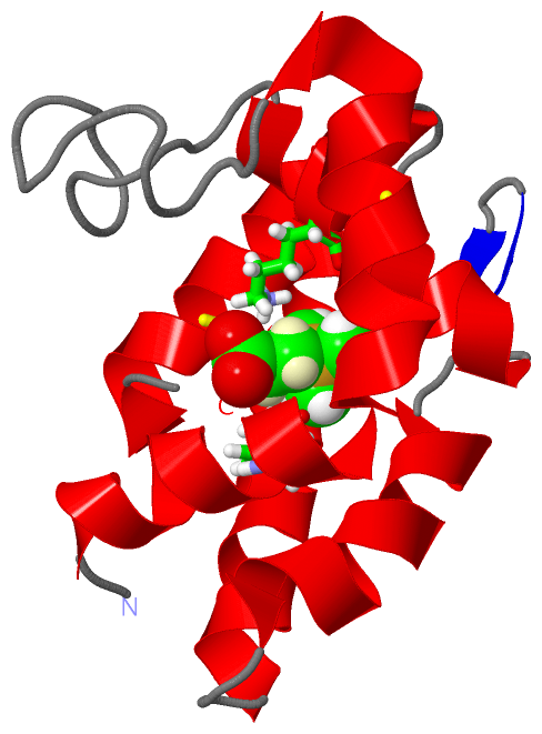
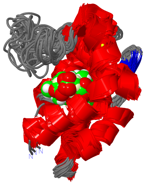
NMR Structure (1, 1)
|
Sites (1, 1)
NMR Structure (1, 1)
|
SS Bonds (3, 3)
NMR Structure
|
||||||||||||||||
Cis Peptide Bonds (0, 0)| (no "Cis Peptide Bond" information available for 1TUJ) |
SAPs(SNPs)/Variants (0, 0)| (no "SAP(SNP)/Variant" information available for 1TUJ) |
PROSITE Motifs (0, 0)| (no "PROSITE Motif" information available for 1TUJ) |
Exons (0, 0)| (no "Exon" information available for 1TUJ) |
Sequences/Alignments
NMR StructureChain A from PDB Type:PROTEIN Length:123 aligned with Q9U9J5_APIME | Q9U9J5 from UniProtKB/TrEMBL Length:142 Alignment length:123 29 39 49 59 69 79 89 99 109 119 129 139 Q9U9J5_APIME 20 IDQDTVVAKYMEYLMPDIMPCADELHISEDIATNIQAAKNGADMSQLGCLKACVMKRIEMLKGTELYVEPVYKMIEVVHAGNADDIQLVKGIANECIENAKGETDECNIGNKYTDCYIEKLFS 142 SCOP domains --------------------------------------------------------------------------------------------------------------------------- SCOP domains CATH domains --------------------------------------------------------------------------------------------------------------------------- CATH domains Pfam domains PBP_GOBP-1tujA01 A:1-121 -- Pfam domains SAPs(SNPs) --------------------------------------------------------------------------------------------------------------------------- SAPs(SNPs) PROSITE --------------------------------------------------------------------------------------------------------------------------- PROSITE Transcript --------------------------------------------------------------------------------------------------------------------------- Transcript 1tuj A 1 IDQDTVVAKYMEYLMPDIMPCADELHISEDIATNIQAAKNGADMSQLGCLKACVMKRIEMLKGTELYVEPVYKMIEVVHAGNADDIQLVKGIANECIENAKGETDECNIGNKYTDCYIEKLFS 123 10 20 30 40 50 60 70 80 90 100 110 120
|
||||||||||||||||||||
SCOP Domains (0, 0)| (no "SCOP Domain" information available for 1TUJ) |
CATH Domains (0, 0)| (no "CATH Domain" information available for 1TUJ) |
Pfam Domains (1, 1)
NMR Structure
|
Gene Ontology (1, 1)|
NMR Structure(hide GO term definitions) Chain A (Q9U9J5_APIME | Q9U9J5)
|
||||||||||||
Interactive Views
|
||||||||||||||||||||||||||||||||||||||||||||||||||||||||||||||||||||||||||||||||||||||||||||||||||||||||||||||||||||||
Still Images
|
||||||||||||||||
Databases
|
||||||||||||||||||||||||||||||||||||||||||||||||||||||||||||||||||||||||||||||||||||||||||||||||||||||||||||||||||||||||||||||||||||||||||||||||||||||||||||||||
Analysis Tools
|
|||||||||||||||||||||||||||||||||||||||||||||||||||||||||||||
Entries Sharing at Least One Protein Chain (UniProt ID)
Related Entries Specified in the PDB File
|
|