Show PDB file:   
         Plain Text   HTML   (compressed file size)
QuickSearch:   
by PDB,NDB,UniProt,PROSITE Code or Search Term(s)  
(-)Asymmetric Unit
(-)Asym. Unit - sites
(-)Biological Unit 1
(-)Biol. Unit 1 - sites
(-)Biological Unit 2
(-)Biological Unit 3
collapse expand < >
Image Asymmetric Unit
Asymmetric Unit  (Jmol Viewer)
Image Asym. Unit - sites
Asym. Unit - sites  (Jmol Viewer)
Image Biological Unit 1
Biological Unit 1  (Jmol Viewer)
Image Biol. Unit 1 - sites
Biol. Unit 1 - sites  (Jmol Viewer)
Image Biological Unit 2
Biological Unit 2  (Jmol Viewer)
Image Biological Unit 3
Biological Unit 3  (Jmol Viewer)

(-) Description

Title :  CRYSTAL STRUCTURE OF PFAL009167 A PUTATIVE D-RIBULOSE 5-PHOSPHATE 3-EPIMERASE FROM P.FALCIPARUM
 
Authors :  J. Caruthers, J. Bosch, W. G. J. Hol, Structural Genomics Of Pathoge Protozoa Consortium (Sgpp)
Date :  18 Jun 04  (Deposition) - 21 Dec 04  (Release) - 13 Jul 11  (Revision)
Method :  X-RAY DIFFRACTION
Resolution :  2.00
Chains :  Asym. Unit :  A,B
Biol. Unit 1:  A  (1x)
Biol. Unit 2:  B  (1x)
Biol. Unit 3:  A (1x),B (1x)
Keywords :  Structural Genomics, Protein Structure Initiative, Psi, Structural Genomics Of Pathogenic Protozoa Consortium, Sgpp, Tim Barrel, Ribulose Phosphate Epimerase, Unknown Function (Keyword Search: [Gene Ontology, PubMed, Web (Google))
 
Reference :  J. Caruthers, J. Bosch, F. Buckner, W. Van Voorhis, P. Myler, E. Worthey, C. Mehlin, E. Boni, G. Detitta, J. Luft, A. Lauricella, O. Kalyuzhniy, L. Anderson, F. Zucker, M. Soltis, W. G. Hol
Structure Of A Ribulose 5-Phosphate 3-Epimerase From Plasmodium Falciparum.
Proteins V. 62 338 2006
PubMed-ID: 16304640  |  Reference-DOI: 10.1002/PROT.20764

(-) Compounds

Molecule 1 - D-RIBULOSE-5-PHOSPHATE 3-EPIMERASE, PUTATIVE
    ChainsA, B
    EngineeredYES
    Expression SystemESCHERICHIA COLI BL21
    Expression System PlasmidLIC
    Expression System StrainBL21
    Expression System Taxid511693
    Expression System Vector TypePLASMID
    Organism ScientificPLASMODIUM FALCIPARUM
    Organism Taxid36329
    Strain3D7

 Structural Features

(-) Chains, Units

  12
Asymmetric Unit AB
Biological Unit 1 (1x)A 
Biological Unit 2 (1x) B
Biological Unit 3 (1x)A (1x)B (1x)

Summary Information (see also Sequences/Alignments below)

(-) Ligands, Modified Residues, Ions  (2, 4)

Asymmetric Unit (2, 4)
No.NameCountTypeFull Name
1SO42Ligand/IonSULFATE ION
2ZN2Ligand/IonZINC ION
Biological Unit 1 (1, 1)
No.NameCountTypeFull Name
1SO41Ligand/IonSULFATE ION
2ZN-1Ligand/IonZINC ION
Biological Unit 2 (1, 1)
No.NameCountTypeFull Name
1SO41Ligand/IonSULFATE ION
2ZN-1Ligand/IonZINC ION
Biological Unit 3 (1, 1)
No.NameCountTypeFull Name
1SO41Ligand/IonSULFATE ION
2ZN-1Ligand/IonZINC ION

(-) Sites  (4, 4)

Asymmetric Unit (4, 4)
No.NameEvidenceResiduesDescription
1AC1SOFTWAREGLY A:152 , GLY A:153 , GLY A:181 , GLY A:201 , THR A:202 , HOH A:1001 , HOH A:1019 , HOH A:1033 , HOH A:1039 , HOH A:1232BINDING SITE FOR RESIDUE SO4 A 2224
2AC2SOFTWAREGLY B:152 , GLY B:153 , GLY B:180 , GLY B:181 , GLY B:201 , THR B:202 , HOH B:1055 , HOH B:1073 , HOH B:1087 , HOH B:1093 , HOH B:1239BINDING SITE FOR RESIDUE SO4 B 3224
3AC3SOFTWAREHIS A:36 , ASP A:38 , HIS A:70 , ASP A:179BINDING SITE FOR RESIDUE ZN A 900
4AC4SOFTWAREHIS B:36 , ASP B:38 , HIS B:70 , ASP B:179BINDING SITE FOR RESIDUE ZN B 902

(-) SS Bonds  (0, 0)

(no "SS Bond" information available for 1TQX)

(-) Cis Peptide Bonds  (0, 0)

(no "Cis Peptide Bond" information available for 1TQX)

 Sequence-Structure Mapping

(-) SAPs(SNPs)/Variants  (0, 0)

(no "SAP(SNP)/Variant" information available for 1TQX)

(-) PROSITE Motifs  (0, 0)

(no "PROSITE Motif" information available for 1TQX)

(-) Exons   (0, 0)

(no "Exon" information available for 1TQX)

(-) Sequences/Alignments

Asymmetric Unit
   Reformat: Number of residues per line =  ('0' or empty: single-line sequence representation)
  Number of residues per labelling interval =   
  UniProt sequence: complete  aligned part    
   Show mapping: SCOP domains CATH domains Pfam domains Secondary structure (by author)
SAPs(SNPs) PROSITE motifs Exons
(details for a mapped element are shown in a popup box when the mouse pointer rests over it)
Chain A from PDB  Type:PROTEIN  Length:221
 aligned with Q8I5L3_PLAF7 | Q8I5L3 from UniProtKB/TrEMBL  Length:227

    Alignment length:221
                                    13        23        33        43        53        63        73        83        93       103       113       123       133       143       153       163       173       183       193       203       213       223 
         Q8I5L3_PLAF7     4 LKAIIAPSVLASNISKLAEETQRMESLGAEWIHLDVMDMHFVPNLSFGPPVINNLKKYTKSIFFDVHLMVEYPEKYVPLLKTSNQLTFHFEALNEDTERCIQLAKEIRDNNLWCGISIKPKTDVQKLVPILDTNLINTVLVMTVEPGFGGQSFMHDMMGKVSFLRKKYKNLNIQVDGGLNIETTEISASHGANIIVAGTSIFNAEDPKYVIDTMRVSVQKY 224
               SCOP domains d1tqxa_ A: D-ribulose-5-phosphate 3-epimerase                                                                                                                                                                                 SCOP domains
               CATH domains 1tqxA00 A:4-224 Aldolase class I                                                                                                                                                                                              CATH domains
               Pfam domains ----------------------------------------------------------------------------------------------------------------------------------------------------------------------------------------------------------------------------- Pfam domains
         Sec.struct. author ...eeee.hhhhhhhhhhhhhhhhhhh...eeeeeee..........hhhhhhhhhhhh...eeeeeee..hhhhhhhhh....eeeee.hhhh.hhhhhhhhhhhhhh...eeeeee....hhhhhhhhhh.....eeeee...........hhhhhhhhhhhhhhh...eeeee...hhhhhhhhhhh...eeeehhhhhh..hhhhhhhhhhhhhhhh Sec.struct. author
                 SAPs(SNPs) ----------------------------------------------------------------------------------------------------------------------------------------------------------------------------------------------------------------------------- SAPs(SNPs)
                    PROSITE ----------------------------------------------------------------------------------------------------------------------------------------------------------------------------------------------------------------------------- PROSITE
                 Transcript ----------------------------------------------------------------------------------------------------------------------------------------------------------------------------------------------------------------------------- Transcript
                 1tqx A   4 LKAIIAPSVLASNISKLAEETQRMESLGAEWIHLDVMDMHFVPNLSFGPPVINNLKKYTKSIFFDVHLMVEYPEKYVPLLKTSNQLTFHFEALNEDTERCIQLAKEIRDNNLWCGISIKPKTDVQKLVPILDTNLINTVLVMTVEPGFGGQSFMHDMMGKVSFLRKKYKNLNIQVDGGLNIETTEISASHGANIIVAGTSIFNAEDPKYVIDTMRVSVQKY 224
                                    13        23        33        43        53        63        73        83        93       103       113       123       133       143       153       163       173       183       193       203       213       223 

Chain B from PDB  Type:PROTEIN  Length:221
 aligned with Q8I5L3_PLAF7 | Q8I5L3 from UniProtKB/TrEMBL  Length:227

    Alignment length:221
                                    13        23        33        43        53        63        73        83        93       103       113       123       133       143       153       163       173       183       193       203       213       223 
         Q8I5L3_PLAF7     4 LKAIIAPSVLASNISKLAEETQRMESLGAEWIHLDVMDMHFVPNLSFGPPVINNLKKYTKSIFFDVHLMVEYPEKYVPLLKTSNQLTFHFEALNEDTERCIQLAKEIRDNNLWCGISIKPKTDVQKLVPILDTNLINTVLVMTVEPGFGGQSFMHDMMGKVSFLRKKYKNLNIQVDGGLNIETTEISASHGANIIVAGTSIFNAEDPKYVIDTMRVSVQKY 224
               SCOP domains d1tqxb_ B: D-ribulose-5-phosphate 3-epimerase                                                                                                                                                                                 SCOP domains
               CATH domains 1tqxB00 B:4-224 Aldolase class I                                                                                                                                                                                              CATH domains
           Pfam domains (1) ---Ribul_P_3_epim-1tqxB01 B:7-208                                                                                                                                                                            ---------------- Pfam domains (1)
           Pfam domains (2) ---Ribul_P_3_epim-1tqxB02 B:7-208                                                                                                                                                                            ---------------- Pfam domains (2)
         Sec.struct. author ...eeee.hhhhhhhhhhhhhhhhhhh...eeeeeee..........hhhhhhhhhhhh...eeeeeee..hhhhhhhhh....eeeee.hhhh.hhhhhhhhhhhhhhh..eeeeee....hhhhhhhhhh.....eeeee...........hhhhhhhhhhhhhhh...eeeee...hhhhhhhhhhh...eeeehhhhhh..hhhhhhhhhhhhhhh. Sec.struct. author
                 SAPs(SNPs) ----------------------------------------------------------------------------------------------------------------------------------------------------------------------------------------------------------------------------- SAPs(SNPs)
                    PROSITE ----------------------------------------------------------------------------------------------------------------------------------------------------------------------------------------------------------------------------- PROSITE
                 Transcript ----------------------------------------------------------------------------------------------------------------------------------------------------------------------------------------------------------------------------- Transcript
                 1tqx B   4 LKAIIAPSVLASNISKLAEETQRMESLGAEWIHLDVMDMHFVPNLSFGPPVINNLKKYTKSIFFDVHLMVEYPEKYVPLLKTSNQLTFHFEALNEDTERCIQLAKEIRDNNLWCGISIKPKTDVQKLVPILDTNLINTVLVMTVEPGFGGQSFMHDMMGKVSFLRKKYKNLNIQVDGGLNIETTEISASHGANIIVAGTSIFNAEDPKYVIDTMRVSVQKY 224
                                    13        23        33        43        53        63        73        83        93       103       113       123       133       143       153       163       173       183       193       203       213       223 

   Legend:   → Mismatch (orange background)
  - → Gap (green background, '-', border residues have a numbering label)
    → Modified Residue (blue background, lower-case, 'x' indicates undefined single-letter code, labelled with number + name)
  x → Chemical Group (purple background, 'x', labelled with number + name, e.g. ACE or NH2)
  extra numbering lines below/above indicate numbering irregularities and modified residue names etc., number ends below/above '|'

 Classification and Annotation

(-) SCOP Domains  (1, 2)

Asymmetric Unit

(-) CATH Domains  (1, 2)

Asymmetric Unit
(-)
Class: Alpha Beta (26913)

(-) Pfam Domains  (1, 2)

Asymmetric Unit

(-) Gene Ontology  (12, 12)

Asymmetric Unit(hide GO term definitions)
Chain A,B   (Q8I5L3_PLAF7 | Q8I5L3)
molecular function
    GO:0003824    catalytic activity    Catalysis of a biochemical reaction at physiological temperatures. In biologically catalyzed reactions, the reactants are known as substrates, and the catalysts are naturally occurring macromolecular substances known as enzymes. Enzymes possess specific binding sites for substrates, and are usually composed wholly or largely of protein, but RNA that has catalytic activity (ribozyme) is often also regarded as enzymatic.
    GO:0016853    isomerase activity    Catalysis of the geometric or structural changes within one molecule. Isomerase is the systematic name for any enzyme of EC class 5.
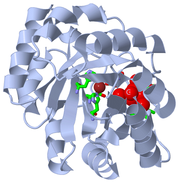
    GO:0046872    metal ion binding    Interacting selectively and non-covalently with any metal ion.
    GO:0016857    racemase and epimerase activity, acting on carbohydrates and derivatives    Catalysis of a reaction that alters the configuration of one or more chiral centers in a carbohydrate molecule.
    GO:0004750    ribulose-phosphate 3-epimerase activity    Catalysis of the reaction: D-ribulose 5-phosphate = D-xylulose 5-phosphate.
biological process
    GO:0005975    carbohydrate metabolic process    The chemical reactions and pathways involving carbohydrates, any of a group of organic compounds based of the general formula Cx(H2O)y. Includes the formation of carbohydrate derivatives by the addition of a carbohydrate residue to another molecule.
    GO:0044262    cellular carbohydrate metabolic process    The chemical reactions and pathways involving carbohydrates, any of a group of organic compounds based of the general formula Cx(H2O)y, as carried out by individual cells.
    GO:0008152    metabolic process    The chemical reactions and pathways, including anabolism and catabolism, by which living organisms transform chemical substances. Metabolic processes typically transform small molecules, but also include macromolecular processes such as DNA repair and replication, and protein synthesis and degradation.
    GO:0019323    pentose catabolic process    The chemical reactions and pathways resulting in the breakdown of a pentose, any monosaccharide with a chain of five carbon atoms in the molecule.
    GO:0006098    pentose-phosphate shunt    The glucose-6-phosphate catabolic process in which, coupled to NADPH synthesis, glucose-6-P is oxidized with the formation of carbon dioxide (CO2) and ribulose 5-phosphate; ribulose 5-P then enters a series of reactions interconverting sugar phosphates. The pentose phosphate pathway is a major source of reducing equivalents for biosynthesis reactions and is also important for the conversion of hexoses to pentoses.
    GO:0009052    pentose-phosphate shunt, non-oxidative branch    The branch of the pentose-phosphate shunt which does not involve oxidation reactions. It comprises a series of sugar phosphate interconversions, starting with ribulose 5-P and producing fructose 6-P and glyceraldehyde 3-P.
cellular component
    GO:0005829    cytosol    The part of the cytoplasm that does not contain organelles but which does contain other particulate matter, such as protein complexes.

 Visualization

(-) Interactive Views

Asymmetric Unit
  Complete Structure
    Jena3D(integrated viewing of ligand, site, SAP, PROSITE, SCOP information)
    WebMol | AstexViewer[tm]@PDBe
(Java Applets, require no local installation except for Java; loading may be slow)
    STRAP
(Java WebStart application, automatic local installation, requires Java; full application with system access!)
    RasMol
(require local installation)
    Molscript (VRML)
(requires installation of a VRML viewer; select preferred view via VRML and generate a mono or stereo PDF format file)
 
  Ligands, Modified Residues, Ions
    SO4  [ RasMol | Jena3D ]  +environment [ RasMol | Jena3D ]
    ZN  [ RasMol | Jena3D ]  +environment [ RasMol | Jena3D ]
 
  Sites
    AC1  [ RasMol ]  +environment [ RasMol ]
    AC2  [ RasMol ]  +environment [ RasMol ]
    AC3  [ RasMol ]  +environment [ RasMol ]
    AC4  [ RasMol ]  +environment [ RasMol ]
 
  Cis Peptide Bonds
(no "Cis Peptide Bonds" information available for 1tqx)
 
Biological Units
  Complete Structure
    Biological Unit 1  [ Jena3D ]
    Biological Unit 2  [ Jena3D ]
    Biological Unit 3  [ Jena3D ]

(-) Still Images

Jmol
  protein: cartoon or spacefill or dots and stick; nucleic acid: cartoon and stick; ligands: spacefill; active site: stick
Molscript
  protein, nucleic acid: cartoon; ligands: spacefill; active site: ball and stick

 Databases and Analysis Tools

(-) Databases

Access by PDB/NDB ID
  1tqx
    Family and Domain InformationProDom | SYSTERS
    General Structural InformationGlycoscienceDB | MMDB | NDB | OCA | PDB | PDBe | PDBj | PDBsum | PDBWiki | PQS | PROTEOPEDIA
    Orientation in MembranesOPM
    Protein SurfaceSURFACE
    Secondary StructureDSSP (structure derived) | HSSP (homology derived)
    Structural GenomicsGeneCensus
    Structural NeighboursCE | VAST
    Structure ClassificationCATH | Dali | SCOP
    Validation and Original DataBMRB Data View | BMRB Restraints Grid | EDS | PROCHECK | RECOORD | WHAT_CHECK
 
Access by UniProt ID/Accession number
  Q8I5L3_PLAF7 | Q8I5L3
    Comparative Protein Structure ModelsModBase
    Genomic InformationEnsembl
    Protein-protein InteractionDIP
    Sequence, Family and Domain InformationInterPro | Pfam | SMART | UniProtKB/TrEMBL
 
Access by Enzyme Classificator   (EC Number)
  (no 'Enzyme Classificator' available)
    General Enzyme InformationBRENDA | EC-PDB | Enzyme | IntEnz
    PathwayKEGG | MetaCyc
 
Access by Disease Identifier   (MIM ID)
  (no 'MIM ID' available)
    Disease InformationOMIM
 
Access by GenAge ID
  (no 'GenAge ID' available)
    Age Related InformationGenAge

(-) Analysis Tools

Access by PDB/NDB ID
    Domain InformationXDom
    Interatomic Contacts of Structural UnitsCSU
    Ligand-protein ContactsLPC
    Protein CavitiescastP
    Sequence and Secondary StructurePDBCartoon
    Structure AlignmentSTRAP(Java WebStart application, automatic local installation, requires Java; full application with system access!)
    Structure and Sequence BrowserSTING
 
Access by UniProt ID/Accession number
  Q8I5L3_PLAF7 | Q8I5L3
    Protein Disorder PredictionDisEMBL | FoldIndex | GLOBPLOT (for more information see DisProt)

 Related Entries

(-) Entries Sharing at Least One Protein Chain (UniProt ID)

(no "Entries Sharing at Least One Protein Chain" available for 1TQX)

(-) Related Entries Specified in the PDB File

(no "Related Entries Specified in the PDB File" available for 1TQX)