|
|
|
|
 Description
|
|
Compounds
|
||||||||||||||||||||||||||||||||||||||||||||
Chains, Units
Summary Information (see also Sequences/Alignments below) |
Ligands, Modified Residues, Ions (0, 0)| (no "Ligand,Modified Residues,Ions" information available for 1T3B) |
Sites (0, 0)| (no "Site" information available for 1T3B) |
SS Bonds (1, 1)
Asymmetric Unit
|
||||||||
Cis Peptide Bonds (1, 1)
Asymmetric Unit
|
||||||||
SAPs(SNPs)/Variants (0, 0)| (no "SAP(SNP)/Variant" information available for 1T3B) |
PROSITE Motifs (0, 0)| (no "PROSITE Motif" information available for 1T3B) |
Exons (0, 0)| (no "Exon" information available for 1T3B) |
Sequences/Alignments
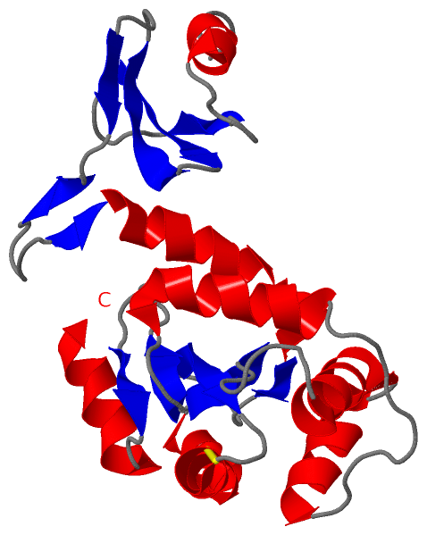
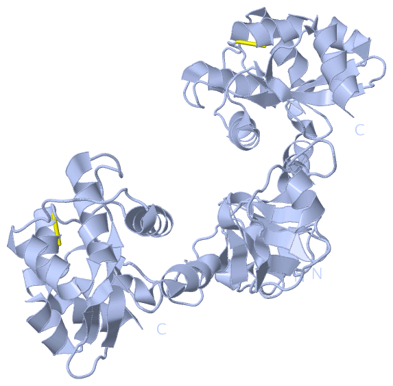
Asymmetric UnitChain A from PDB Type:PROTEIN Length:209 aligned with DSBC_HAEIN | P45111 from UniProtKB/Swiss-Prot Length:229 Alignment length:209 29 39 49 59 69 79 89 99 109 119 129 139 149 159 169 179 189 199 209 219 DSBC_HAEIN 20 DAAIKRKLQSFNISNIVIKSSPISGIKTAVTDQGILYVSEDGKYLFEGKLYELTNNGPVDVAGKILVDKLNSYKDEMIVYPAKNEKHVVTVFMDITCHYCHLLHQQLKEYNDLGITVRYLAFPRAGMNNQTAKQMEAIWTAKDPVFALNEAEKGNLPKEVKTPNIVKKHYELGIQFGVRGTPSIVTSTGELIGGYLKPADLLRALEETA 228 SCOP domains d1t3ba2 A:2-60 d1t3ba1 A:61-210 Disulfide bond isomerase, DsbC, C-terminal domain SCOP domains CATH domains 1t3bA01 A:2-49 1t3bA02 A:50-210 Glutaredoxin CATH domains Pfam domains ---DsbC_N-1t3bA02 A:5-58 ----------------------Thioredoxin_2-1t3bA01 A:81-206 ---- Pfam domains SAPs(SNPs) ----------------------------------------------------------------------------------------------------------------------------------------------------------------------------------------------------------------- SAPs(SNPs) PROSITE ----------------------------------------------------------------------------------------------------------------------------------------------------------------------------------------------------------------- PROSITE Transcript ----------------------------------------------------------------------------------------------------------------------------------------------------------------------------------------------------------------- Transcript 1t3b A 2 DAAIKRKLQSFNISNIVIKSSPISGIKTAVTDQGILYVSEDGKYLFEGKLYELTNNGPVDVAGKILVDKLNSYKDEMIVYPAKNEKHVVTVFMDITCHYCHLLHQQLKEYNDLGITVRYLAFPRAGMNNQTAKQMEAIWTAKDPVFALNEAEKGNLPKEVKTPNIVKKHYELGIQFGVRGTPSIVTSTGELIGGYLKPADLLRALEETA 210 11 21 31 41 51 61 71 81 91 101 111 121 131 141 151 161 171 181 191 201
|
||||||||||||||||||||
SCOP Domains (2, 2)
Asymmetric Unit
 |
CATH Domains (2, 2)
Asymmetric Unit
|
Pfam Domains (2, 2)| Asymmetric Unit |
Gene Ontology (1, 1)|
Asymmetric Unit(hide GO term definitions) Chain A (DSBC_HAEIN | P45111)
|
||||||||||||
Interactive Views
|
|||||||||||||||||||||||||||||||||||||||||||||||||||||||||||||||||||||||||||||||||||||||||||||||||||||||||||||||||||||||||||||||||||||||
Still Images
|
||||||||||||||||
Databases
|
||||||||||||||||||||||||||||||||||||||||||||||||||||||||||||||||||||||||||||||||||||||||||||||||||||||||||||||||||||||||||||||||||||||||||||||||||||||||||||||||
Analysis Tools
|
|||||||||||||||||||||||||||||||||||||||||||||||||||||||||||||
Entries Sharing at Least One Protein Chain (UniProt ID)
Related Entries Specified in the PDB File
|
|