|
|
|
|
 Description
|
|
Compounds
|
||||||||||||||||||||||||||||||||||||||||||||
Chains, Units
Summary Information (see also Sequences/Alignments below) |
Ligands, Modified Residues, Ions (2, 3)| Asymmetric/Biological Unit (2, 3) |
Sites (3, 3)
Asymmetric Unit (3, 3)
|
SS Bonds (0, 0)| (no "SS Bond" information available for 1T1G) |
Cis Peptide Bonds (3, 3)
Asymmetric/Biological Unit
|
||||||||||||||||
SAPs(SNPs)/Variants (0, 0)| (no "SAP(SNP)/Variant" information available for 1T1G) |
PROSITE Motifs (0, 0)| (no "PROSITE Motif" information available for 1T1G) |
Exons (0, 0)| (no "Exon" information available for 1T1G) |
Sequences/Alignments
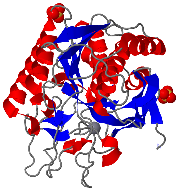
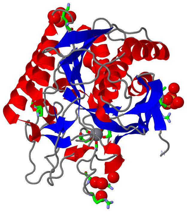
Asymmetric/Biological UnitChain A from PDB Type:PROTEIN Length:357 aligned with Q8RR56_9BACI | Q8RR56 from UniProtKB/TrEMBL Length:552 Alignment length:357 198 208 218 228 238 248 258 268 278 288 298 308 318 328 338 348 358 368 378 388 398 408 418 428 438 448 458 468 478 488 498 508 518 528 538 Q8RR56_9BACI 189 AAPTAYTPLDVAQAYQFPEGLDGQGQCIAIIELGGGYDETSLAQYFASLGVSAPQVVSVSVDGATNQPTGDPNGPDGEVELDIEVAGALAPGAKIAVYFAPNTDAGFLNAITTAVHDPTHKPSIVSISWGGPEDSWAPASIAAMNRAFLDAAALGVTVLAAAGDSGSTDGEQDGLYHVDFPAASPYVLACGGTRLVASAGRIERETVWNDGPDGGSTGGGVSRIFPLPSWQERANVPPSANPGAGSGRGVPDVAGNADPATGYEVVIDGETTVIGGTSAVAPLFAALVARINQKLGKPVGYLNPTLYQLPPEVFHDITEGNNDIANRARIYQAGPGWDPCTGLGSPIGIRLLQALLP 545 SCOP domains d1t1ga_ A: Serine-carboxyl proteinase, SCP SCOP domains CATH domains 1t1gA00 A:1-357 [code=3.40.50.200, no name defined] CATH domains Pfam domains ---------------------------------------------------------------------------Peptidase_S8-1t1gA01 A:76-331 -------------------------- Pfam domains SAPs(SNPs) --------------------------------------------------------------------------------------------------------------------------------------------------------------------------------------------------------------------------------------------------------------------------------------------------------------------------------------------------------------------- SAPs(SNPs) PROSITE --------------------------------------------------------------------------------------------------------------------------------------------------------------------------------------------------------------------------------------------------------------------------------------------------------------------------------------------------------------------- PROSITE Transcript --------------------------------------------------------------------------------------------------------------------------------------------------------------------------------------------------------------------------------------------------------------------------------------------------------------------------------------------------------------------- Transcript 1t1g A 1 AAPTAYTPLDVAQAYQFPEGLDGQGQCIAIIALGGGYDETSLAQYFASLGVSAPQVVSVSVDGATNQPTGDPNGPDGEVELDIEVAGALAPGAKIAVYFAPNTDAGFLNAITTAVHDPTHKPSIVSISWGGPEDSWAPASIAAMNRAFLDAAALGVTVLAAAGDSGSTDGEQDGLYHVDFPAASPYVLACGGTRLVASAGRIERETVWNDGPDGGSTGGGVSRIFPLPSWQERANVPPSANPGAGSGRGVPDVAGNADPATGYEVVIDGETTVIGGTSAVAPLFAALVARINQKLGKPVGYLNPTLYQLPPEVFHDITEGNNDIANRARIYQAGPGWDPCTGLGSPIGIRLLQALLP 357 10 20 30 40 50 60 70 80 90 100 110 120 130 140 150 160 170 180 190 200 210 220 230 240 250 260 270 280 290 300 310 320 330 340 350
|
||||||||||||||||||||
SCOP Domains (1, 1)
Asymmetric/Biological Unit
 |
CATH Domains (1, 1)
Asymmetric/Biological Unit
|
Pfam Domains (1, 1)
Asymmetric/Biological Unit
|
Gene Ontology (4, 4)|
Asymmetric/Biological Unit(hide GO term definitions) Chain A (Q8RR56_9BACI | Q8RR56)
|
||||||||||||||||||||||||||||||||||||
Interactive Views
|
||||||||||||||||||||||||||||||||||||||||||||||||||||||||||||||||||||||||||||||||||||||||||||||||||||||||||||||||||||||||||||||||||||||||||||||||||||||||||
Still Images
|
||||||||||||||||
Databases
|
||||||||||||||||||||||||||||||||||||||||||||||||||||||||||||||||||||||||||||||||||||||||||||||||||||||||||||||||||||||||||||||||||||||||||||||||||||||||||||||||
Analysis Tools
|
|||||||||||||||||||||||||||||||||||||||||||||||||||||||||||||
Entries Sharing at Least One Protein Chain (UniProt ID)
Related Entries Specified in the PDB File
|
|