Show PDB file:   
         Plain Text   HTML   (compressed file size)
QuickSearch:   
by PDB,NDB,UniProt,PROSITE Code or Search Term(s)  
(-)Asym.Unit - manually
(-)Asymmetric Unit
(-)Asym. Unit - sites
(-)Biological Unit 1
(-)Biol. Unit 1 - sites
(-)Biological Unit 2
collapse expand < >
Image Asym.Unit - manually
Asym.Unit - manually  (Jmol Viewer)
Image Asymmetric Unit
Asymmetric Unit  (Jmol Viewer)
Image Asym. Unit - sites
Asym. Unit - sites  (Jmol Viewer)
Image Biological Unit 1
Biological Unit 1  (Jmol Viewer)
Image Biol. Unit 1 - sites
Biol. Unit 1 - sites  (Jmol Viewer)
Image Biological Unit 2
Biological Unit 2  (Jmol Viewer)

(-) Description

Title :  CRYSTAL STRUCTURE OF THE HUMAN MBL-ASSOCIATED PROTEIN 19 (MAP19)
 
Authors :  L. A. Gregory, N. M. Thielens, G. J. Arlaud, J. C. Fontecilla-Camps, C. Gaboriaud
Date :  05 Apr 04  (Deposition) - 22 Jun 04  (Release) - 24 Feb 09  (Revision)
Method :  X-RAY DIFFRACTION
Resolution :  2.50
Chains :  Asym. Unit :  A,B
Biol. Unit 1:  A,B  (1x)
Biol. Unit 2:  A,B  (2x)
Keywords :  Calcium, Complement, Innate Immunity, Cub, Egf, Protein Binding (Keyword Search: [Gene Ontology, PubMed, Web (Google))
 
Reference :  L. A. Gregory, N. M. Thielens, M. Matsushita, R. Sorensen, G. J. Arlaud, J. C. Fontecilla-Camps, C. Gaboriaud
The X-Ray Structure Of Human Mbl-Associated Protein 19 (Map19) And Its Interaction Site With Mannan-Binding Lectin And L-Ficolin
J. Biol. Chem. V. 279 29391 2004
PubMed-ID: 15117939  |  Reference-DOI: 10.1074/JBC.M402687200
(for further references see the PDB file header)

(-) Compounds

Molecule 1 - MANNOSE BINDING LECTIN-ASSOCIATED SERINE PROTEASE-2 RELATED PROTEIN, MAP19 (19KDA)
    ChainsA, B
    EngineeredYES
    Expression SystemTRICHOPLUSIA NI
    Expression System CommonCABBAGE LOOPER
    Expression System StrainHIGH FIVE INSECT CELLS
    Expression System Taxid7111
    Expression System VectorPBC1S
    Expression System Vector TypeBACULOVIRUS
    GeneMASP2
    Organism CommonHUMAN
    Organism ScientificHOMO SAPIENS
    Organism Taxid9606

 Structural Features

(-) Chains, Units

  12
Asymmetric Unit AB
Biological Unit 1 (1x)AB
Biological Unit 2 (2x)AB

Summary Information (see also Sequences/Alignments below)

(-) Ligands, Modified Residues, Ions  (1, 4)

Asymmetric Unit (1, 4)
No.NameCountTypeFull Name
1CA4Ligand/IonCALCIUM ION
Biological Unit 1 (0, 0)
No.NameCountTypeFull Name
1CA-1Ligand/IonCALCIUM ION
Biological Unit 2 (0, 0)
No.NameCountTypeFull Name
1CA-1Ligand/IonCALCIUM ION

(-) Sites  (4, 4)

Asymmetric Unit (4, 4)
No.NameEvidenceResiduesDescription
1AC1SOFTWAREASP A:123 , ILE A:124 , GLU A:126 , ASN A:143 , HIS A:144 , GLY A:147 , HOH A:1009BINDING SITE FOR RESIDUE CA A 1001
2AC2SOFTWAREASP B:123 , ILE B:124 , GLU B:126 , ASN B:143 , HIS B:144 , GLY B:147 , HOH B:1007BINDING SITE FOR RESIDUE CA B 1002
3AC3SOFTWAREGLU A:52 , ASP A:60 , ASP A:105 , SER A:107 , ASN A:108 , HOH A:1019BINDING SITE FOR RESIDUE CA A 1003
4AC4SOFTWAREGLU B:52 , ASP B:60 , ASP B:105 , SER B:107 , ASN B:108BINDING SITE FOR RESIDUE CA B 1004

(-) SS Bonds  (8, 8)

Asymmetric Unit
No.Residues
1A:57 -A:75
2A:127 -A:141
3A:137 -A:150
4A:152 -A:165
5B:57 -B:75
6B:127 -B:141
7B:137 -B:150
8B:152 -B:165

(-) Cis Peptide Bonds  (2, 2)

Asymmetric Unit
No.Residues
1Phe A:20 -Pro A:21
2Phe B:20 -Pro B:21

 Sequence-Structure Mapping

(-) SAPs(SNPs)/Variants  (6, 12)

Asymmetric Unit (6, 12)
  dbSNPPDB
No.SourceVariant IDVariantUniProt IDStatusIDChainVariant
1UniProtVAR_025344R99QMASP2_HUMANPolymorphism61735600A/BR84Q
2UniProtVAR_025345R118CMASP2_HUMANPolymorphism147270785A/BR103C
3UniProtVAR_025346D120GMASP2_HUMANDisease (MASPD)72550870A/BD105G
4UniProtVAR_025347P126LMASP2_HUMANDisease (MASPD)56392418A/BP111L
5UniProtVAR_075087T128MMASP2_HUMANPolymorphism141145402A/BT113M
6UniProtVAR_028784H155RMASP2_HUMANPolymorphism2273343A/BH140R

  SNP/SAP Summary Statistics (UniProtKB/Swiss-Prot)
Biological Unit 1 (6, 12)
  dbSNPPDB
No.SourceVariant IDVariantUniProt IDStatusIDChainVariant
1UniProtVAR_025344R99QMASP2_HUMANPolymorphism61735600A/BR84Q
2UniProtVAR_025345R118CMASP2_HUMANPolymorphism147270785A/BR103C
3UniProtVAR_025346D120GMASP2_HUMANDisease (MASPD)72550870A/BD105G
4UniProtVAR_025347P126LMASP2_HUMANDisease (MASPD)56392418A/BP111L
5UniProtVAR_075087T128MMASP2_HUMANPolymorphism141145402A/BT113M
6UniProtVAR_028784H155RMASP2_HUMANPolymorphism2273343A/BH140R

  SNP/SAP Summary Statistics (UniProtKB/Swiss-Prot)
Biological Unit 2 (6, 24)
  dbSNPPDB
No.SourceVariant IDVariantUniProt IDStatusIDChainVariant
1UniProtVAR_025344R99QMASP2_HUMANPolymorphism61735600A/BR84Q
2UniProtVAR_025345R118CMASP2_HUMANPolymorphism147270785A/BR103C
3UniProtVAR_025346D120GMASP2_HUMANDisease (MASPD)72550870A/BD105G
4UniProtVAR_025347P126LMASP2_HUMANDisease (MASPD)56392418A/BP111L
5UniProtVAR_075087T128MMASP2_HUMANPolymorphism141145402A/BT113M
6UniProtVAR_028784H155RMASP2_HUMANPolymorphism2273343A/BH140R

  SNP/SAP Summary Statistics (UniProtKB/Swiss-Prot)

(-) PROSITE Motifs  (3, 6)

Asymmetric Unit (3, 6)
 PROSITEUniProtKBPDB
No.IDACDescriptionIDLocationCountLocation
1EGF_CAPS01187 Calcium-binding EGF-like domain signature.MASP2_HUMAN138-165
 
  2A:123-150
B:123-150
2ASX_HYDROXYLPS00010 Aspartic acid and asparagine hydroxylation site.MASP2_HUMAN156-167
 
  2A:141-152
B:141-152
3EGF_2PS01186 EGF-like domain signature 2.MASP2_HUMAN165-180
 
  2A:150-165
B:150-165
Biological Unit 1 (3, 6)
 PROSITEUniProtKBPDB
No.IDACDescriptionIDLocationCountLocation
1EGF_CAPS01187 Calcium-binding EGF-like domain signature.MASP2_HUMAN138-165
 
  2A:123-150
B:123-150
2ASX_HYDROXYLPS00010 Aspartic acid and asparagine hydroxylation site.MASP2_HUMAN156-167
 
  2A:141-152
B:141-152
3EGF_2PS01186 EGF-like domain signature 2.MASP2_HUMAN165-180
 
  2A:150-165
B:150-165
Biological Unit 2 (3, 12)
 PROSITEUniProtKBPDB
No.IDACDescriptionIDLocationCountLocation
1EGF_CAPS01187 Calcium-binding EGF-like domain signature.MASP2_HUMAN138-165
 
  4A:123-150
B:123-150
2ASX_HYDROXYLPS00010 Aspartic acid and asparagine hydroxylation site.MASP2_HUMAN156-167
 
  4A:141-152
B:141-152
3EGF_2PS01186 EGF-like domain signature 2.MASP2_HUMAN165-180
 
  4A:150-165
B:150-165

(-) Exons   (4, 8)

Asymmetric Unit (4, 8)
 ENSEMBLUniProtKBPDB
No.Transcript IDExonExon IDGenome LocationLengthIDLocationLengthCountLocationLength
1.1dENST000004008971dENSE00001833377chr1:11107280-1110726021MASP2_HUMAN1-220--
1.2ENST000004008972ENSE00000818831chr1:11107176-11106948229MASP2_HUMAN2-78772A:2-63
B:3-63
62
61
1.3aENST000004008973aENSE00000472116chr1:11106790-11106613178MASP2_HUMAN79-138602A:64-123
B:64-123
60
60
1.3cENST000004008973cENSE00000743093chr1:11105596-11105465132MASP2_HUMAN138-182452A:123-167
B:123-167
45
45
1.4ENST000004008974ENSE00000472118chr1:11103592-11103396197MASP2_HUMAN182-247662A:167-168
B:167-168
2
2
1.5bENST000004008975bENSE00000743090chr1:11103079-11102932148MASP2_HUMAN248-297500--
1.6ENST000004008976ENSE00000472120chr1:11097868-11097750119MASP2_HUMAN297-336400--
1.7ENST000004008977ENSE00000743085chr1:11094963-1109488579MASP2_HUMAN337-363270--
1.8ENST000004008978ENSE00000743084chr1:11090939-11090805135MASP2_HUMAN363-408460--
1.9ENST000004008979ENSE00000472123chr1:11090307-1109023375MASP2_HUMAN408-433260--
1.10ENST0000040089710ENSE00000818830chr1:11087705-110865801126MASP2_HUMAN433-6862540--

(-) Sequences/Alignments

Asymmetric Unit
   Reformat: Number of residues per line =  ('0' or empty: single-line sequence representation)
  Number of residues per labelling interval =   
  UniProt sequence: complete  aligned part    
   Show mapping: SCOP domains CATH domains Pfam domains Secondary structure (by author)
SAPs(SNPs) PROSITE motifs Exons
(details for a mapped element are shown in a popup box when the mouse pointer rests over it)
Chain A from PDB  Type:PROTEIN  Length:167
 aligned with MASP2_HUMAN | O00187 from UniProtKB/Swiss-Prot  Length:686

    Alignment length:167
                                    26        36        46        56        66        76        86        96       106       116       126       136       146       156       166       176       
          MASP2_HUMAN    17 PLGPKWPEPVFGRLASPGFPGEYANDQERRWTLTAPPGYRLRLYFTHFDLELSHLCEYDFVKLSSGAKVLATLCGQESTDTERAPGKDTFYSLGSSLDITFRSDYSNEKPFTGFEAFYAAEDIDECQVAPGEAPTCDHHCHNHLGGFYCSCRAGYVLHRNKRTCSAL 183
               SCOP domains -d1szba1 A:3-123 Mannose-binding protein associated serine protease 2, MASP2                                              d1szba2 A:124-168                             SCOP domains
               CATH domains 1szbA01 A:2-125  [code=2.60.120.290, no name defined]                                                                       1szbA02 A:126-168 Laminin                   CATH domains
               Pfam domains ----------------------------------------------------------------------------------------------------------------------------------------------------------------------- Pfam domains
         Sec.struct. author ..........eeeee............eeeeeee....eeeeeeeeeee..........eeeee....eeeee................ee....eeeeeee..........eeeeeeeeee................eeeee..eeeee....eee......eee. Sec.struct. author
                 SAPs(SNPs) ----------------------------------------------------------------------------------Q------------------C-G-----L-M--------------------------R---------------------------- SAPs(SNPs)
                PROSITE (1) -------------------------------------------------------------------------------------------------------------------------EGF_CA  PDB: A:123-150      ------------------ PROSITE (1)
                PROSITE (2) -------------------------------------------------------------------------------------------------------------------------------------------ASX_HYDROXYL---------------- PROSITE (2)
                PROSITE (3) ----------------------------------------------------------------------------------------------------------------------------------------------------EGF_2           --- PROSITE (3)
           Transcript 1 (1) Exon 1.2  PDB: A:2-63 UniProt: 2-78 [INCOMPLETE]              Exon 1.3a  PDB: A:64-123 UniProt: 79-138                    -------------------------------------------1. Transcript 1 (1)
           Transcript 1 (2) -------------------------------------------------------------------------------------------------------------------------Exon 1.3c  PDB: A:123-167 UniProt: 138-182   - Transcript 1 (2)
                 1szb A   2 PLGPKWPEPVFGRLASPGFPGEYANDQERRWTLTAPPGYRLRLYFTHFDLELSHLCEYDFVKLSSGAKVLATLCGQESTDTERAPGKDTFYSLGSSLDITFRSDYSNEKPFTGFEAFYAAEDIDECQVAPGEAPTCDHHCHNHLGGFYCSCRAGYVLHRNKRTCSEQ 168
                                    11        21        31        41        51        61        71        81        91       101       111       121       131       141       151       161       

Chain B from PDB  Type:PROTEIN  Length:166
 aligned with MASP2_HUMAN | O00187 from UniProtKB/Swiss-Prot  Length:686

    Alignment length:166
                                    27        37        47        57        67        77        87        97       107       117       127       137       147       157       167       177      
          MASP2_HUMAN    18 LGPKWPEPVFGRLASPGFPGEYANDQERRWTLTAPPGYRLRLYFTHFDLELSHLCEYDFVKLSSGAKVLATLCGQESTDTERAPGKDTFYSLGSSLDITFRSDYSNEKPFTGFEAFYAAEDIDECQVAPGEAPTCDHHCHNHLGGFYCSCRAGYVLHRNKRTCSAL 183
               SCOP domains d1szbb1 B:3-123 Mannose-binding protein associated serine protease 2, MASP2                                              d1szbb2 B:124-168                             SCOP domains
               CATH domains 1szbB01 B:3-125  [code=2.60.120.290, no name defined]                                                                      1szbB02 B:126-168 Laminin                   CATH domains
           Pfam domains (1) ------CUB-1szbB01 B:9-119                                                                                            ---EGF_CA-1szbB03 B:123-165                   --- Pfam domains (1)
           Pfam domains (2) ------CUB-1szbB02 B:9-119                                                                                            ---EGF_CA-1szbB04 B:123-165                   --- Pfam domains (2)
         Sec.struct. author .........eeeee............eeeeeee.....eeeeeeeeee..........eeeeee..eeeeee................ee....eeeeeee..........eeeeeeeee.................eeeee..eeeee....eee......eee. Sec.struct. author
                 SAPs(SNPs) ---------------------------------------------------------------------------------Q------------------C-G-----L-M--------------------------R---------------------------- SAPs(SNPs)
                PROSITE (1) ------------------------------------------------------------------------------------------------------------------------EGF_CA  PDB: B:123-150      ------------------ PROSITE (1)
                PROSITE (2) ------------------------------------------------------------------------------------------------------------------------------------------ASX_HYDROXYL---------------- PROSITE (2)
                PROSITE (3) ---------------------------------------------------------------------------------------------------------------------------------------------------EGF_2           --- PROSITE (3)
           Transcript 1 (1) Exon 1.2  PDB: B:3-63 UniProt: 2-78 [INCOMPLETE]             Exon 1.3a  PDB: B:64-123 UniProt: 79-138                    -------------------------------------------1. Transcript 1 (1)
           Transcript 1 (2) ------------------------------------------------------------------------------------------------------------------------Exon 1.3c  PDB: B:123-167 UniProt: 138-182   - Transcript 1 (2)
                 1szb B   3 LGPKWPEPVFGRLASPGFPGEYANDQERRWTLTAPPGYRLRLYFTHFDLELSHLCEYDFVKLSSGAKVLATLCGQESTDTERAPGKDTFYSLGSSLDITFRSDYSNEKPFTGFEAFYAAEDIDECQVAPGEAPTCDHHCHNHLGGFYCSCRAGYVLHRNKRTCSEQ 168
                                    12        22        32        42        52        62        72        82        92       102       112       122       132       142       152       162      

   Legend:   → Mismatch (orange background)
  - → Gap (green background, '-', border residues have a numbering label)
    → Modified Residue (blue background, lower-case, 'x' indicates undefined single-letter code, labelled with number + name)
  x → Chemical Group (purple background, 'x', labelled with number + name, e.g. ACE or NH2)
  extra numbering lines below/above indicate numbering irregularities and modified residue names etc., number ends below/above '|'

 Classification and Annotation

(-) SCOP Domains  (2, 4)

Asymmetric Unit

(-) CATH Domains  (2, 4)

Asymmetric Unit
(-)
Class: Mainly Beta (13760)

(-) Pfam Domains  (2, 4)

Asymmetric Unit
(-)
Clan: CUB (11)
(-)
Family: CUB (11)
(-)
Clan: EGF (60)

(-) Gene Ontology  (17, 17)

Asymmetric Unit(hide GO term definitions)
Chain A,B   (MASP2_HUMAN | O00187)
molecular function
    GO:0005509    calcium ion binding    Interacting selectively and non-covalently with calcium ions (Ca2+).
    GO:0048306    calcium-dependent protein binding    Interacting selectively and non-covalently with any protein or protein complex (a complex of two or more proteins that may include other nonprotein molecules), in the presence of calcium.
    GO:0001855    complement component C4b binding    Interacting selectively and non-covalently with the C4b product of the classical complement cascade.
    GO:0016787    hydrolase activity    Catalysis of the hydrolysis of various bonds, e.g. C-O, C-N, C-C, phosphoric anhydride bonds, etc. Hydrolase is the systematic name for any enzyme of EC class 3.
    GO:0046872    metal ion binding    Interacting selectively and non-covalently with any metal ion.
    GO:0008233    peptidase activity    Catalysis of the hydrolysis of a peptide bond. A peptide bond is a covalent bond formed when the carbon atom from the carboxyl group of one amino acid shares electrons with the nitrogen atom from the amino group of a second amino acid.
    GO:0005515    protein binding    Interacting selectively and non-covalently with any protein or protein complex (a complex of two or more proteins that may include other nonprotein molecules).
    GO:0004252    serine-type endopeptidase activity    Catalysis of the hydrolysis of internal, alpha-peptide bonds in a polypeptide chain by a catalytic mechanism that involves a catalytic triad consisting of a serine nucleophile that is activated by a proton relay involving an acidic residue (e.g. aspartate or glutamate) and a basic residue (usually histidine).
    GO:0008236    serine-type peptidase activity    Catalysis of the hydrolysis of peptide bonds in a polypeptide chain by a catalytic mechanism that involves a catalytic triad consisting of a serine nucleophile that is activated by a proton relay involving an acidic residue (e.g. aspartate or glutamate) and a basic residue (usually histidine).
biological process
    GO:0006956    complement activation    Any process involved in the activation of any of the steps of the complement cascade, which allows for the direct killing of microbes, the disposal of immune complexes, and the regulation of other immune processes; the initial steps of complement activation involve one of three pathways, the classical pathway, the alternative pathway, and the lectin pathway, all of which lead to the terminal complement pathway.
    GO:0006958    complement activation, classical pathway    Any process involved in the activation of any of the steps of the classical pathway of the complement cascade which allows for the direct killing of microbes, the disposal of immune complexes, and the regulation of other immune processes.
    GO:0001867    complement activation, lectin pathway    Any process involved in the activation of any of the steps of the lectin pathway of the complement cascade which allows for the direct killing of microbes and the regulation of other immune processes.
    GO:0002376    immune system process    Any process involved in the development or functioning of the immune system, an organismal system for calibrated responses to potential internal or invasive threats.
    GO:0045087    innate immune response    Innate immune responses are defense responses mediated by germline encoded components that directly recognize components of potential pathogens.
    GO:0006508    proteolysis    The hydrolysis of proteins into smaller polypeptides and/or amino acids by cleavage of their peptide bonds.
cellular component
    GO:0070062    extracellular exosome    A vesicle that is released into the extracellular region by fusion of the limiting endosomal membrane of a multivesicular body with the plasma membrane. Extracellular exosomes, also simply called exosomes, have a diameter of about 40-100 nm.
    GO:0005576    extracellular region    The space external to the outermost structure of a cell. For cells without external protective or external encapsulating structures this refers to space outside of the plasma membrane. This term covers the host cell environment outside an intracellular parasite.
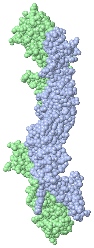
 Visualization

(-) Interactive Views

Asymmetric Unit
  Complete Structure
    Jena3D(integrated viewing of ligand, site, SAP, PROSITE, SCOP information)
    WebMol | AstexViewer[tm]@PDBe
(Java Applets, require no local installation except for Java; loading may be slow)
    STRAP
(Java WebStart application, automatic local installation, requires Java; full application with system access!)
    RasMol
(require local installation)
    Molscript (VRML)
(requires installation of a VRML viewer; select preferred view via VRML and generate a mono or stereo PDF format file)
 
  Ligands, Modified Residues, Ions
    CA  [ RasMol | Jena3D ]  +environment [ RasMol | Jena3D ]
 
  Sites
    AC1  [ RasMol ]  +environment [ RasMol ]
    AC2  [ RasMol ]  +environment [ RasMol ]
    AC3  [ RasMol ]  +environment [ RasMol ]
    AC4  [ RasMol ]  +environment [ RasMol ]
 
  Cis Peptide Bonds
    Phe A:20 - Pro A:21   [ RasMol ]  
    Phe B:20 - Pro B:21   [ RasMol ]  
 
Biological Units
  Complete Structure
    Biological Unit 1  [ Jena3D ]
    Biological Unit 2  [ Jena3D ]

(-) Still Images

Jmol
  protein: cartoon or spacefill or dots and stick; nucleic acid: cartoon and stick; ligands: spacefill; active site: stick
Molscript
  protein, nucleic acid: cartoon; ligands: spacefill; active site: ball and stick

 Databases and Analysis Tools

(-) Databases

Access by PDB/NDB ID
  1szb
    Family and Domain InformationProDom | SYSTERS
    General Structural InformationGlycoscienceDB | MMDB | NDB | OCA | PDB | PDBe | PDBj | PDBsum | PDBWiki | PQS | PROTEOPEDIA
    Orientation in MembranesOPM
    Protein SurfaceSURFACE
    Secondary StructureDSSP (structure derived) | HSSP (homology derived)
    Structural GenomicsGeneCensus
    Structural NeighboursCE | VAST
    Structure ClassificationCATH | Dali | SCOP
    Validation and Original DataBMRB Data View | BMRB Restraints Grid | EDS | PROCHECK | RECOORD | WHAT_CHECK
 
Access by UniProt ID/Accession number
  MASP2_HUMAN | O00187
    Comparative Protein Structure ModelsModBase
    Genomic InformationEnsembl
    Protein-protein InteractionDIP
    Sequence, Family and Domain InformationInterPro | Pfam | SMART | UniProtKB/SwissProt
 
Access by Enzyme Classificator   (EC Number)
  (no 'Enzyme Classificator' available)
    General Enzyme InformationBRENDA | EC-PDB | Enzyme | IntEnz
    PathwayKEGG | MetaCyc
 
Access by Disease Identifier   (MIM ID)
  613791
    Disease InformationOMIM
 
Access by GenAge ID
  (no 'GenAge ID' available)
    Age Related InformationGenAge

(-) Analysis Tools

Access by PDB/NDB ID
    Domain InformationXDom
    Interatomic Contacts of Structural UnitsCSU
    Ligand-protein ContactsLPC
    Protein CavitiescastP
    Sequence and Secondary StructurePDBCartoon
    Structure AlignmentSTRAP(Java WebStart application, automatic local installation, requires Java; full application with system access!)
    Structure and Sequence BrowserSTING
 
Access by UniProt ID/Accession number
  MASP2_HUMAN | O00187
    Protein Disorder PredictionDisEMBL | FoldIndex | GLOBPLOT (for more information see DisProt)

 Related Entries

(-) Entries Sharing at Least One Protein Chain (UniProt ID)

UniProtKB/Swiss-Prot
        MASP2_HUMAN | O001871q3x 1zjk 3tvj 4fxg 5jpm

(-) Related Entries Specified in the PDB File

(no "Related Entries Specified in the PDB File" available for 1SZB)