|
|
|
|
 Description
|
|
Compounds
|
||||||||||||||||||||||||||||||||||||||||||||||||||||
Chains, Units
Summary Information (see also Sequences/Alignments below) |
Ligands, Modified Residues, Ions (0, 0)| (no "Ligand,Modified Residues,Ions" information available for 1SPX) |
Sites (0, 0)| (no "Site" information available for 1SPX) |
SS Bonds (0, 0)| (no "SS Bond" information available for 1SPX) |
Cis Peptide Bonds (0, 0)| (no "Cis Peptide Bond" information available for 1SPX) |
SAPs(SNPs)/Variants (0, 0)| (no "SAP(SNP)/Variant" information available for 1SPX) |
PROSITE Motifs (0, 0)| (no "PROSITE Motif" information available for 1SPX) |
Exons (0, 0)| (no "Exon" information available for 1SPX) |
Sequences/Alignments
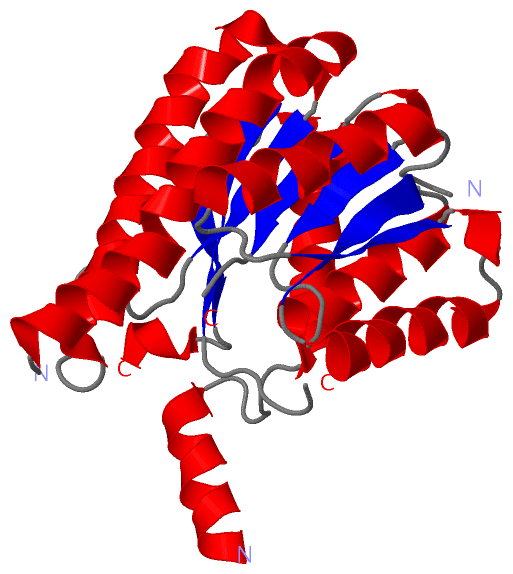
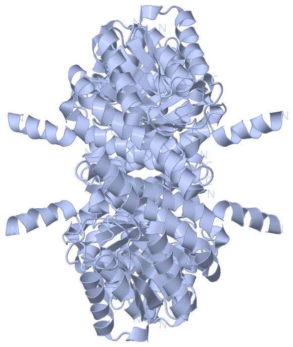
Asymmetric UnitChain A from PDB Type:PROTEIN Length:237 aligned with Q18946_CAEEL | Q18946 from UniProtKB/TrEMBL Length:278 Alignment length:264 11 21 31 41 51 61 71 81 91 101 111 121 131 141 151 161 171 181 191 201 211 221 231 241 251 261 Q18946_CAEEL 2 TRFAEKVAIITGSSNGIGRATAVLFAREGAKVTITGRHAERLEETRQQILAAGVSEQNVNSVVADVTTDAGQDEILSTTLGKFGKLDILVNNAGAAIPDSQSKTGTAQSIESYDATLNLNLRSVIALTKKAVPHLSSTKGEIVNISSIASGLHATPDFPYYSIAKAAIDQYTRNTAIDLIQHGIRVNSISPGLVATGFGSAMGMPEETSKKFYSTMATMKECVPAGVMGQPQDIAEVIAFLADRKTSSYIIGHQLVVDGGSSLI 265 SCOP domains d1spxa_ A: Glucose dehydrogenase (5l265) SCOP domains CATH domains 1spxA00 A:2-265 NAD(P)-binding Rossmann-like Domain CATH domains Pfam domains ----------adh_short_C2-1spxA01 A:12-263 -- Pfam domains SAPs(SNPs) ------------------------------------------------------------------------------------------------------------------------------------------------------------------------------------------------------------------------------------------------------------------------ SAPs(SNPs) PROSITE ------------------------------------------------------------------------------------------------------------------------------------------------------------------------------------------------------------------------------------------------------------------------ PROSITE Transcript ------------------------------------------------------------------------------------------------------------------------------------------------------------------------------------------------------------------------------------------------------------------------ Transcript 1spx A 2 TRFAEKVAIITGSSNGIGRATAVLFAREGAKVTITGRHAERLEETRQQILAAGVSEQNVNSVVADVTTDAGQDEILSTTLGKFGKLDILVNNAG-------------QSIESYDATLNLNLRSVIALTKKAVPHLSSTKGEIVNISSIASGLHATPDFPYYSIAKAAIDQYTRNTAIDLIQHGIRVNSISPGLVATG--------------FYSTMATMKECVPAGVMGQPQDIAEVIAFLADRKTSSYIIGHQLVVDGGSSLI 265 11 21 31 41 51 61 71 81 91 | - 111 121 131 141 151 161 171 181 191 | - - | 221 231 241 251 261 95 109 198 213
|
||||||||||||||||||||
SCOP Domains (1, 1)
Asymmetric Unit
 |
CATH Domains (1, 1)
Asymmetric Unit
|
Pfam Domains (1, 1)
Asymmetric Unit
|
Gene Ontology (2, 2)|
Asymmetric Unit(hide GO term definitions) Chain A (Q18946_CAEEL | Q18946)
|
||||||||||||||||||||||||
Interactive Views
|
||||||||||||||||||||||||||||||||||||||||||||||||||||||||||||||||||||||||||||||||||||||||||||||||||||||||||||||||||||||||||||||||||||||
Still Images
|
||||||||||||||||
Databases
|
||||||||||||||||||||||||||||||||||||||||||||||||||||||||||||||||||||||||||||||||||||||||||||||||||||||||||||||||||||||||||||||||||||||||||||||||||||||||||||||||
Analysis Tools
|
|||||||||||||||||||||||||||||||||||||||||||||||||||||||||||||
Entries Sharing at Least One Protein Chain (UniProt ID)
Related Entries Specified in the PDB File
|
|