|
|
|
|
 Description
|
|
Compounds
|
||||||||||||||||||||||||||||||||||||||||
Chains, Units
Summary Information (see also Sequences/Alignments below) |
Ligands, Modified Residues, Ions (2, 2)| Asymmetric/Biological Unit (2, 2) |
Sites (1, 1)
Asymmetric Unit (1, 1)
|
SS Bonds (0, 0)| (no "SS Bond" information available for 1SPV) |
Cis Peptide Bonds (1, 1)
Asymmetric/Biological Unit
|
||||||||
SAPs(SNPs)/Variants (0, 0)| (no "SAP(SNP)/Variant" information available for 1SPV) |
PROSITE Motifs (0, 0)| (no "PROSITE Motif" information available for 1SPV) |
Exons (0, 0)| (no "Exon" information available for 1SPV) |
Sequences/Alignments
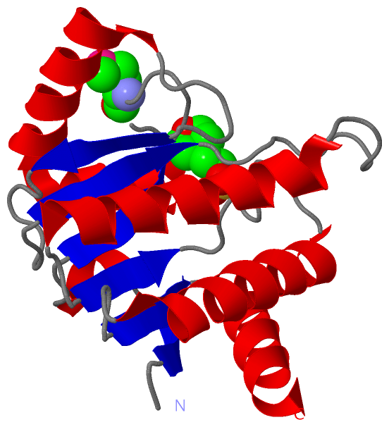
Asymmetric/Biological UnitChain A from PDB Type:PROTEIN Length:172 aligned with YMDB_ECOLI | P0A8D6 from UniProtKB/Swiss-Prot Length:177 Alignment length:172 12 22 32 42 52 62 72 82 92 102 112 122 132 142 152 162 172 YMDB_ECOLI 3 TRIHVVQGDITKLAVDVIVNAANPSLMGGGGVDGAIHRAAGPALLDACLKVRQQQGDCPTGHAVITLAGDLPAKAVVHTVGPVWRGGEQNEDQLLQDAYLNSLRLVAANSYTSVAFPAISTGVYGYPRAAAAEIAVKTVSEFITRHALPEQVYFVCYDEENAHLYERLLTQQ 174 SCOP domains d1spva_ A: Hypothetical protein YmbD SCOP domains CATH domains 1spvA00 A:3-174 Leucine Aminopeptidase, subunit E, domain 1 CATH domains Pfam domains ------------------Macro-1spvA01 A:21-137 ------------------------------------- Pfam domains SAPs(SNPs) ---------------------------------------------------------------------------------------------------------------------------------------------------------------------------- SAPs(SNPs) PROSITE ---------------------------------------------------------------------------------------------------------------------------------------------------------------------------- PROSITE Transcript ---------------------------------------------------------------------------------------------------------------------------------------------------------------------------- Transcript 1spv A 3 TRIHVVQGDITKLAVDVIVNAANPSLmGGGGVDGAIHRAAGPALLDACLKVRQQQGDCPTGHAVITLAGDLPAKAVVHTVGPVWRGGEQNEDQLLQDAYLNSLRLVAANSYTSVAFPAISTGVYGYPRAAAAEIAVKTVSEFITRHALPEQVYFVCYDEENAHLYERLLTQQ 174 12 22 | 32 42 52 62 72 82 92 102 112 122 132 142 152 162 172 29-MSE
|
||||||||||||||||||||
SCOP Domains (1, 1)
Asymmetric/Biological Unit
 |
CATH Domains (1, 1)
Asymmetric/Biological Unit
|
Pfam Domains (1, 1)
Asymmetric/Biological Unit
|
Gene Ontology (10, 10)|
Asymmetric/Biological Unit(hide GO term definitions) Chain A (YMDB_ECOLI | P0A8D6)
|
||||||||||||||||||||||||||||||||||||||||||||||||||||||||||||||||||||||||
Interactive Views
|
||||||||||||||||||||||||||||||||||||||||||||||||||||||||||||||||||||||||||||||||||||||||||||||||||||||||||||||||||||||||||||||
Still Images
|
||||||||||||||||
Databases
|
||||||||||||||||||||||||||||||||||||||||||||||||||||||||||||||||||||||||||||||||||||||||||||||||||||||||||||||||||||||||||||||||||||||||||||||||||||||||||||||||
Analysis Tools
|
|||||||||||||||||||||||||||||||||||||||||||||||||||||||||||||
Entries Sharing at Least One Protein Chain (UniProt ID)
Related Entries Specified in the PDB File
|
|