|
|
|
|
 Description
|
|
Compounds
|
||||||||||||||||||||||||||||||||||||||||||||||||
Chains, Units
Summary Information (see also Sequences/Alignments below) |
Ligands, Modified Residues, Ions (0, 0)| (no "Ligand,Modified Residues,Ions" information available for 1SOU) |
Sites (0, 0)| (no "Site" information available for 1SOU) |
SS Bonds (0, 0)| (no "SS Bond" information available for 1SOU) |
Cis Peptide Bonds (0, 0)| (no "Cis Peptide Bond" information available for 1SOU) |
SAPs(SNPs)/Variants (0, 0)| (no "SAP(SNP)/Variant" information available for 1SOU) |
PROSITE Motifs (0, 0)| (no "PROSITE Motif" information available for 1SOU) |
Exons (0, 0)| (no "Exon" information available for 1SOU) |
Sequences/Alignments
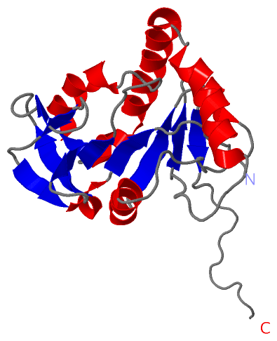
NMR StructureChain A from PDB Type:PROTEIN Length:194 aligned with O67621_AQUAE | O67621 from UniProtKB/TrEMBL Length:186 Alignment length:194 186 10 20 30 40 50 60 70 80 90 100 110 120 130 140 150 160 170 180 | - O67621_AQUAE 1 MLKSELRKKVLHKRINLSEEERRRLSEKVISNLKSLPEFKKSKKVALYCPIKGEVDLTPLFPEVLKEKELILPKVEGNEISLYRVHSPACLGVGAFGIMEPVEGERVNPEDVDFIAVPGVAFDLEGYRLGFGKGYYDRLLKRVKGLKVGVAYSFQVFERLPRDAWDIPVDVLVTEKNVRRLRDGRS-------- - SCOP domains d1soua_ A: Hypothetical protein aq_1731 SCOP domains CATH domains 1souA00 A:1-194 NagB/RpiA/CoA transferase-like CATH domains Pfam domains --5-FTHF_cyc-lig-1souA01 A:3-175 ------------------- Pfam domains SAPs(SNPs) -------------------------------------------------------------------------------------------------------------------------------------------------------------------------------------------------- SAPs(SNPs) PROSITE -------------------------------------------------------------------------------------------------------------------------------------------------------------------------------------------------- PROSITE Transcript -------------------------------------------------------------------------------------------------------------------------------------------------------------------------------------------------- Transcript 1sou A 1 MLKSELRKKVLHKRINLSEEERRRLSEKVISNLKSLPEFKKSKKVALYCPIKGEVDLTPLFPEVLKEKELILPKVEGNEISLYRVHSPACLGVGAFGIMEPVEGERVNPEDVDFIAVPGVAFDLEGYRLGFGKGYYDRLLKRVKGLKVGVAYSFQVFERLPRDAWDIPVDVLVTEKNVRRLRDGRSLEHHHHHH 194 10 20 30 40 50 60 70 80 90 100 110 120 130 140 150 160 170 180 190
|
||||||||||||||||||||
SCOP Domains (1, 1)
NMR Structure
 |
CATH Domains (1, 1)
NMR Structure
|
Pfam Domains (1, 1)
NMR Structure
|
Gene Ontology (6, 6)|
NMR Structure(hide GO term definitions) Chain A (O67621_AQUAE | O67621)
|
||||||||||||||||||||||||||||||||||||||||||||||||
Interactive Views
|
||||||||||||||||||||||||||||||||||||||||||||||||||||||||||||||||||||||||||||||||||||||||||||||||||||||||||||||||||||
Still Images
|
||||||||||||||||
Databases
|
||||||||||||||||||||||||||||||||||||||||||||||||||||||||||||||||||||||||||||||||||||||||||||||||||||||||||||||||||||||||||||||||||||||||||||||||||||||||||||||||
Analysis Tools
|
|||||||||||||||||||||||||||||||||||||||||||||||||||||||||||||
Entries Sharing at Least One Protein Chain (UniProt ID)
Related Entries Specified in the PDB File
|
|