|
|
|
|
 Description
|
|
Compounds
|
||||||||||||||||||||||||||||||||||||||||||||||||
Chains, Units
Summary Information (see also Sequences/Alignments below) |
Ligands, Modified Residues, Ions (0, 0)| (no "Ligand,Modified Residues,Ions" information available for 1SF0) |
Sites (0, 0)| (no "Site" information available for 1SF0) |
SS Bonds (0, 0)| (no "SS Bond" information available for 1SF0) |
Cis Peptide Bonds (0, 0)| (no "Cis Peptide Bond" information available for 1SF0) |
SAPs(SNPs)/Variants (0, 0)| (no "SAP(SNP)/Variant" information available for 1SF0) |
PROSITE Motifs (0, 0)| (no "PROSITE Motif" information available for 1SF0) |
Exons (0, 0)| (no "Exon" information available for 1SF0) |
Sequences/Alignments
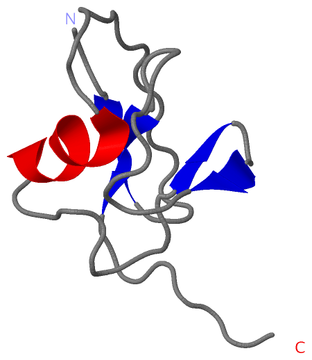
NMR StructureChain A from PDB Type:PROTEIN Length:68 aligned with Q8U1Z3_PYRFU | Q8U1Z3 from UniProtKB/TrEMBL Length:69 Alignment length:68 11 21 31 41 51 61 Q8U1Z3_PYRFU 2 KMIKVKVIGRNIEKEIEWREGMKVRDILRAVGFNTESAIAKVNGKVVLEDDEVKDGDFVEVIPVVSGG 69 SCOP domains d1sf0a_ A: Hypothetical protein PF1061 SCOP domains CATH domains 1sf0A00 A:2-69 [code=3.10.20.30, no name defined] CATH domains Pfam domains ---ThiS-1sf0A01 A:5-69 Pfam domains SAPs(SNPs) -------------------------------------------------------------------- SAPs(SNPs) PROSITE -------------------------------------------------------------------- PROSITE Transcript -------------------------------------------------------------------- Transcript 1sf0 A 2 KMIKVKVIGRNIEKEIEWREGMKVRDILRAVGFNTESAIAKVNGKVVLEDDEVKDGDFVEVIPVVSGG 69 11 21 31 41 51 61
|
||||||||||||||||||||
SCOP Domains (1, 1)
NMR Structure
 |
CATH Domains (1, 1)
NMR Structure
|
Pfam Domains (1, 1)
NMR Structure
|
Gene Ontology (0, 0)|
NMR Structure(hide GO term definitions)
(no "Gene Ontology" information available for 1SF0)
|
Interactive Views
|
||||||||||||||||||||||||||||||||||||||||||||||||||||||||||||||||||||||||||||||||||||||||||||||||||||||||||||||||||||
Still Images
|
||||||||||||||||
Databases
|
||||||||||||||||||||||||||||||||||||||||||||||||||||||||||||||||||||||||||||||||||||||||||||||||||||||||||||||||||||||||||||||||||||||||||||||||||||||||||||||||
Analysis Tools
|
|||||||||||||||||||||||||||||||||||||||||||||||||||||||||||||
Entries Sharing at Least One Protein Chain (UniProt ID)
Related Entries Specified in the PDB File
|
|