|
|
|
|
 Description
|
|
Compounds
|
||||||||||||||||||||||||
Chains, Units
Summary Information (see also Sequences/Alignments below) |
Ligands, Modified Residues, Ions (0, 0)| (no "Ligand,Modified Residues,Ions" information available for 1S6D) |
Sites (0, 0)| (no "Site" information available for 1S6D) |
SS Bonds (4, 4)
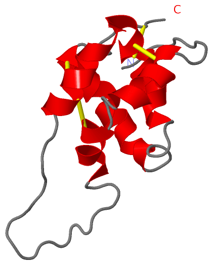
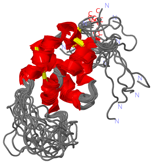
NMR Structure
|
||||||||||||||||||||
Cis Peptide Bonds (0, 0)| (no "Cis Peptide Bond" information available for 1S6D) |
SAPs(SNPs)/Variants (0, 0)| (no "SAP(SNP)/Variant" information available for 1S6D) |
PROSITE Motifs (0, 0)| (no "PROSITE Motif" information available for 1S6D) |
Exons (0, 0)| (no "Exon" information available for 1S6D) |
Sequences/Alignments
NMR StructureChain A from PDB Type:PROTEIN Length:103 aligned with 2SS8_HELAN | P23110 from UniProtKB/Swiss-Prot Length:141 Alignment length:103 48 58 68 78 88 98 108 118 128 138 2SS8_HELAN 39 PYGRGRTESGCYQQMEEAEMLNHCGMYLMKNLGERSQVSPRMREEDHKQLCCMQLKNLDEKCMCPAIMMMLNEPMWIRMRDQVMSMAHNLPIECNLMSQPCQM 141 SCOP domains d1s6da_ A: Methionine-rich 2S protein (albumin 8) SCOP domains CATH domains ---------1s6dA01 A:10-94 Bifunctional Trypsin/Alpha-Amylase Inhibitor --------- CATH domains Pfam domains --------Tryp_alpha_amyl-1s6dA01 A:9-102 - Pfam domains SAPs(SNPs) ------------------------------------------------------------------------------------------------------- SAPs(SNPs) PROSITE ------------------------------------------------------------------------------------------------------- PROSITE Transcript ------------------------------------------------------------------------------------------------------- Transcript 1s6d A 1 PYGRGRTESGCYQQMEEAEMLNHCGMYLMKNLGERSQVSPRMREEDHKQLCCMQLKNLDEKCMCPAIMMMLNEPMWIRMRDQVMSMAHNLPIECNLMSQPCQM 103 10 20 30 40 50 60 70 80 90 100
|
||||||||||||||||||||
SCOP Domains (1, 1)
NMR Structure
 |
CATH Domains (1, 1)
NMR Structure
|
Pfam Domains (1, 1)
NMR Structure
|
Gene Ontology (1, 1)|
NMR Structure(hide GO term definitions) Chain A (2SS8_HELAN | P23110)
|
||||||||||||
Interactive Views
|
||||||||||||||||||||||||||||||||||||||||||||||||||||||||||||||||||||||||||||||||||||||||||||||||||||||||||||||||||||
Still Images
|
||||||||||||||||
Databases
|
||||||||||||||||||||||||||||||||||||||||||||||||||||||||||||||||||||||||||||||||||||||||||||||||||||||||||||||||||||||||||||||||||||||||||||||||||||||||||||||||
Analysis Tools
|
|||||||||||||||||||||||||||||||||||||||||||||||||||||||||||||
Entries Sharing at Least One Protein Chain (UniProt ID)
Related Entries Specified in the PDB File
|
|