|
|
|
|
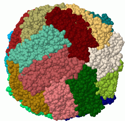
 Description
|
|
Compounds
|
||||||||||||||||||||||||||||||||||||||||||||||||||||||||||||
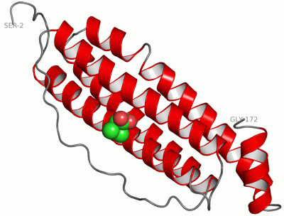
Chains, Units
Summary Information (see also Sequences/Alignments below) |
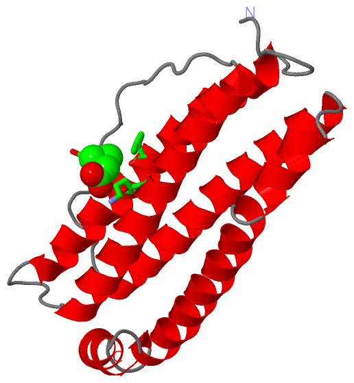
Ligands, Modified Residues, Ions (1, 1)
Asymmetric Unit (1, 1)
|
Sites (1, 1)
Asymmetric Unit (1, 1)
|
SS Bonds (0, 0)| (no "SS Bond" information available for 1RCC) |
Cis Peptide Bonds (1, 1)
Asymmetric Unit
|
||||||||
SAPs(SNPs)/Variants (0, 0)| (no "SAP(SNP)/Variant" information available for 1RCC) |
PROSITE Motifs (3, 3)
Asymmetric Unit (3, 3)
|
||||||||||||||||||||||||||||||||||||||||||||||||||||||||||||||||||||||||||||||||
Exons (0, 0)| (no "Exon" information available for 1RCC) |
Sequences/Alignments
Asymmetric UnitChain A from PDB Type:PROTEIN Length:171 aligned with FRI3_LITCT | P07797 from UniProtKB/Swiss-Prot Length:173 Alignment length:171 12 22 32 42 52 62 72 82 92 102 112 122 132 142 152 162 172 FRI3_LITCT 3 SQVRQNFHQDCEAGLNRTVNLKFHSSYVYLSMASYFNRDDVALSNFAKFFRERSEEEKEHAEKLIEYQNQRGGRVFLQSVEKPERDDWANGLEALQTALKLQKSVNQALLDLHAVAADKSDPHMTDFLESPYLSESVETIKKLGDHITSLKKLWSSHPGMAEYLFNKHTLG 173 SCOP domains d1rcca_ A: (Apo)ferritin SCOP domains CATH domains 1rccA00 A:2-172 [code=1.20.1260.10, no name defined] CATH domains Pfam domains --------------------------------------------------------------------------------------------------------------------------------------------------------------------------- Pfam domains SAPs(SNPs) --------------------------------------------------------------------------------------------------------------------------------------------------------------------------- SAPs(SNPs) PROSITE (1) ----FERRITIN_LIKE PDB: A:6-155 UniProt: 7-156 ----------------- PROSITE (1) PROSITE (2) -------------------------------------------------------FERRITIN_1 ----------------------------------------------FERRITIN_2 ------------------------------ PROSITE (2) Transcript --------------------------------------------------------------------------------------------------------------------------------------------------------------------------- Transcript 1rcc A 2 SQVRQNFHQDCEAGLNRTVNLKFHSSYVYLSMASYFNRDDVALSNFAKFFRERSAAAKAHAEKLIEYQNQRGGRVFLQSVEKPERDDWANGLEALQTALKLQKSVNQALLDLHAVAADKSDPHMTDFLESPYLSESVETIKKLGDHITSLKKLWSSHPGMAEYLFNKHTLG 172 11 21 31 41 51 61 71 81 91 101 111 121 131 141 151 161 171
|
||||||||||||||||||||
SCOP Domains (1, 1)
Asymmetric Unit
 |
CATH Domains (1, 1)
Asymmetric Unit
|
Pfam Domains (0, 0)| (no "Pfam Domain" information available for 1RCC) |
Gene Ontology (5, 5)|
Asymmetric Unit(hide GO term definitions) Chain A (FRI3_LITCT | P07797)
|
||||||||||||||||||||||||||||||||||||||||||||||||
Interactive Views
|
|||||||||||||||||||||||||||||||||||||||||||||||||||||||||||||||||||||||||||||||||||||||||||||||||||||||||||||||||||||||||||||||||||||||||
Still Images
|
||||||||||||||||
Databases
|
||||||||||||||||||||||||||||||||||||||||||||||||||||||||||||||||||||||||||||||||||||||||||||||||||||||||||||||||||||||||||||||||||||||||||||||||||||||||||||||||
Analysis Tools
|
|||||||||||||||||||||||||||||||||||||||||||||||||||||||||||||
Entries Sharing at Least One Protein Chain (UniProt ID)
Related Entries Specified in the PDB File
|
|