|
|
|
|
 Description
|
|
Compounds
|
||||||||||||||||||||||||||||||||||||||||||||||||||||
Chains, Units
Summary Information (see also Sequences/Alignments below) |
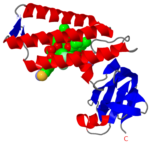
Ligands, Modified Residues, Ions (3, 4)| Asymmetric/Biological Unit (3, 4) |
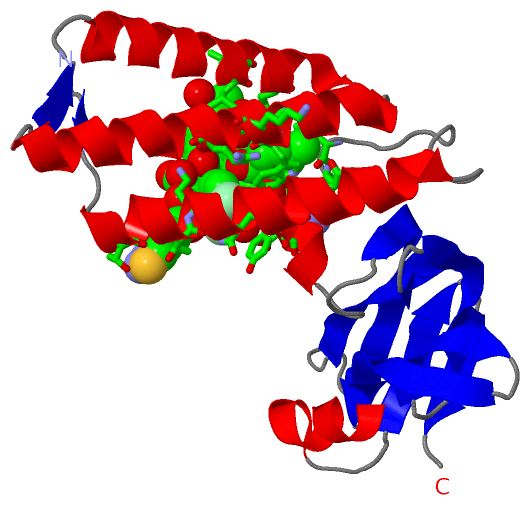
Sites (4, 4)
Asymmetric Unit (4, 4)
|
SS Bonds (0, 0)| (no "SS Bond" information available for 1R6N) |
Cis Peptide Bonds (1, 1)
Asymmetric/Biological Unit
|
||||||||
SAPs(SNPs)/Variants (0, 0)| (no "SAP(SNP)/Variant" information available for 1R6N) |
PROSITE Motifs (0, 0)| (no "PROSITE Motif" information available for 1R6N) |
Exons (0, 0)| (no "Exon" information available for 1R6N) |
Sequences/Alignments
Asymmetric/Biological UnitChain A from PDB Type:PROTEIN Length:196 aligned with VE2_HPV11 | P04015 from UniProtKB/Swiss-Prot Length:367 Alignment length:196 10 20 30 40 50 60 70 80 90 100 110 120 130 140 150 160 170 180 190 VE2_HPV11 1 MEAIAKRLDACQDQLLELYEENSIDIHKHIMHWKCIRLESVLLHKAKQMGLSHIGLQVVPPLTVSETKGHNAIEMQMHLESLAKTQYGVEPWTLQDTSYEMWLTPPKRCFKKQGNTVEVKFDGCEDNVMEYVVWTHIYLQDNDSWVKVTSSVDAKGIYYTCGQFKTYYVNFNKEAQKYGSTNHWEVCYGSTVICSP 196 SCOP domains d1r6na_ A: E2 regulatory, transactivation domain SCOP domains CATH domains 1r6nA01 A:1-98 [code=1.10.287.30, no name defined] 1r6nA02 A:99-196 [code=2.170.200.10, no name defined] CATH domains Pfam domains -PPV_E2_N-1r6nA01 A:2-196 Pfam domains SAPs(SNPs) ---------------------------------------------------------------------------------------------------------------------------------------------------------------------------------------------------- SAPs(SNPs) PROSITE ---------------------------------------------------------------------------------------------------------------------------------------------------------------------------------------------------- PROSITE Transcript ---------------------------------------------------------------------------------------------------------------------------------------------------------------------------------------------------- Transcript 1r6n A 1 HEAIAKRLDACQDQLLELYEENSIDIHKHIMHWKCIRLESVLLHKAKQMGLSHIGLQVVPPLTVSETKGHNAIEMQMHLESLAKTQYGVEPWTLQDTSYEMWLTPPKRCFKKQGNTVEVKFDGCEDNVMEYVVWTHIYLQDNDSWVKVTSSVDAKGIYYTCGQFKTYYVNFNKEAQKYGSTNHWEVCYGSTVICSP 196 10 20 30 40 50 60 70 80 90 100 110 120 130 140 150 160 170 180 190
|
||||||||||||||||||||
SCOP Domains (1, 1)
Asymmetric/Biological Unit
 |
CATH Domains (2, 2)
Asymmetric/Biological Unit
|
Pfam Domains (1, 1)
Asymmetric/Biological Unit
|
Gene Ontology (11, 11)|
Asymmetric/Biological Unit(hide GO term definitions) Chain A (VE2_HPV11 | P04015)
|
||||||||||||||||||||||||||||||||||||||||||||||||||||||||||||||||||||||||||||||||||||
Interactive Views
|
||||||||||||||||||||||||||||||||||||||||||||||||||||||||||||||||||||||||||||||||||||||||||||||||||||||||||||||||||||||||||||||||||||||||||||||||||||||||||
Still Images
|
||||||||||||||||
Databases
|
||||||||||||||||||||||||||||||||||||||||||||||||||||||||||||||||||||||||||||||||||||||||||||||||||||||||||||||||||||||||||||||||||||||||||||||||||||||||||||||||
Analysis Tools
|
|||||||||||||||||||||||||||||||||||||||||||||||||||||||||||||
Entries Sharing at Least One Protein Chain (UniProt ID)
Related Entries Specified in the PDB File
|
|