Show PDB file:   
         Plain Text   HTML   (compressed file size)
QuickSearch:   
by PDB,NDB,UniProt,PROSITE Code or Search Term(s)  
(-)Asymmetric Unit
(-)Asym. Unit - sites
(-)Biological Unit 1
(-)Biol. Unit 1 - sites
collapse expand < >
Image Asymmetric Unit
Asymmetric Unit  (Jmol Viewer)
Image Asym. Unit - sites
Asym. Unit - sites  (Jmol Viewer)
Image Biological Unit 1
Biological Unit 1  (Jmol Viewer)
Image Biol. Unit 1 - sites
Biol. Unit 1 - sites  (Jmol Viewer)

(-) Description

Title :  CRYSTAL STRUCTURE OF METHANOCOCCUS JANNASCHII PHOSPHOSULFOLACTATE SYNTHASE
 
Authors :  E. L. Wise, D. E. Graham, R. H. White, I. Rayment
Date :  02 Sep 03  (Deposition) - 09 Dec 03  (Release) - 13 Jul 11  (Revision)
Method :  X-RAY DIFFRACTION
Resolution :  1.60
Chains :  Asym. Unit :  A
Biol. Unit 1:  A  (3x)
Keywords :  Beta-Alpha-Barrel, Lyase (Keyword Search: [Gene Ontology, PubMed, Web (Google))
 
Reference :  E. L. Wise, D. E. Graham, R. H. White, I. Rayment
The Structural Determination Of Phosphosulfolactate Synthas From Methanococcus Jannaschii At 1. 7-A Resolution: An Enolase That Is Not An Enolase
J. Biol. Chem. V. 278 45858 2003
PubMed-ID: 12952952  |  Reference-DOI: 10.1074/JBC.M307486200

(-) Compounds

Molecule 1 - (2R)-PHOSPHO-3-SULFOLACTATE SYNTHASE
    ChainsA
    EC Number4.4.-.-
    EngineeredYES
    Expression SystemESCHERICHIA COLI
    Expression System StrainBL21 (DE3) CODONPLUS
    Expression System Taxid562
    Expression System Vector TypePLASMID
    GeneCOMA
    Organism ScientificMETHANOCALDOCOCCUS JANNASCHII
    Organism Taxid2190
    SynonymPHOSPHOSULFOLACTATE SYNTHASE;
PSL SYNTHASE

 Structural Features

(-) Chains, Units

  1
Asymmetric Unit A
Biological Unit 1 (3x)A

Summary Information (see also Sequences/Alignments below)

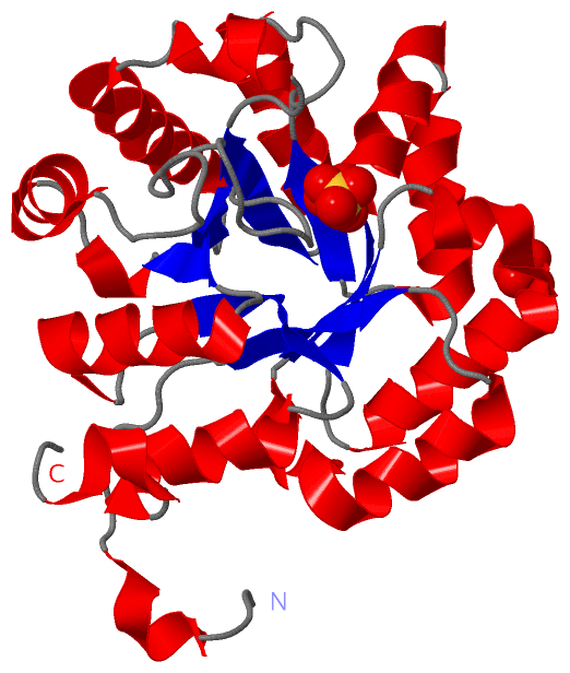
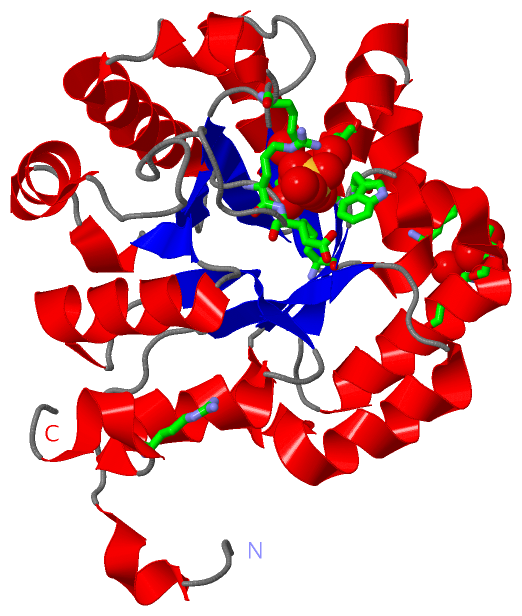
(-) Ligands, Modified Residues, Ions  (1, 3)

Asymmetric Unit (1, 3)
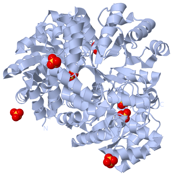
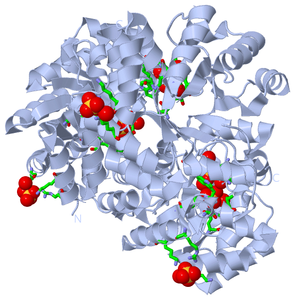
No.NameCountTypeFull Name
1SO43Ligand/IonSULFATE ION
Biological Unit 1 (1, 9)
No.NameCountTypeFull Name
1SO49Ligand/IonSULFATE ION

(-) Sites  (3, 3)

Asymmetric Unit (3, 3)
No.NameEvidenceResiduesDescription
1AC1SOFTWARETRP A:46 , LYS A:137 , ARG A:170 , GLU A:171 , ARG A:244 , HOH A:321 , HOH A:330 , HOH A:353BINDING SITE FOR RESIDUE SO4 A 300
2AC2SOFTWARELYS A:43 , TRP A:46 , GLY A:74 , GLY A:75 , THR A:76 , GLU A:103 , LYS A:137 , HOH A:321 , HOH A:444BINDING SITE FOR RESIDUE SO4 A 301
3AC3SOFTWAREARG A:54 , LYS A:58 , LYS A:96 , HOH A:504BINDING SITE FOR RESIDUE SO4 A 302

(-) SS Bonds  (0, 0)

(no "SS Bond" information available for 1QWG)

(-) Cis Peptide Bonds  (0, 0)

(no "Cis Peptide Bond" information available for 1QWG)

 Sequence-Structure Mapping

(-) SAPs(SNPs)/Variants  (0, 0)

(no "SAP(SNP)/Variant" information available for 1QWG)

(-) PROSITE Motifs  (0, 0)

(no "PROSITE Motif" information available for 1QWG)

(-) Exons   (0, 0)

(no "Exon" information available for 1QWG)

(-) Sequences/Alignments

Asymmetric Unit
   Reformat: Number of residues per line =  ('0' or empty: single-line sequence representation)
  Number of residues per labelling interval =   
  UniProt sequence: complete  aligned part    
   Show mapping: SCOP domains CATH domains Pfam domains Secondary structure (by author)
SAPs(SNPs) PROSITE motifs Exons
(details for a mapped element are shown in a popup box when the mouse pointer rests over it)
Chain A from PDB  Type:PROTEIN  Length:251
 aligned with PSLS_METJA | Q57703 from UniProtKB/Swiss-Prot  Length:251

    Alignment length:251
                                    10        20        30        40        50        60        70        80        90       100       110       120       130       140       150       160       170       180       190       200       210       220       230       240       250 
           PSLS_METJA     1 MKAFEFLYEDFQRGLTVVLDKGLPPKFVEDYLKVCGDYIDFVKFGWGTSAVIDRDVVKEKINYYKDWGIKVYPGGTLFEYAYSKGKFDEFLNECEKLGFEAVEISDGSSDISLEERKNAIKRAKDNGFMVLTEVGKKMPDKDKQLTIDDRIKLINFDLDAGADYVIIEGRESGKGIGLFDKEGKVKENELDVLAKNVDINKVIFEAPQKSQQVAFILKFGSSVNLANIAFDEVISLETLRRGLRGDTFGKV 251
               SCOP domains d1qwga_ A: (2r)-phospho-3-sulfolactate synthase ComA                                                                                                                                                                                                        SCOP domains
               CATH domains 1qwgA00 A:1-251 Aldolase class I                                                                                                                                                                                                                            CATH domains
               Pfam domains ComA-1qwgA01 A:1-246                                                                                                                                                                                                                                  ----- Pfam domains
         Sec.struct. author ...hhhhh.......eeeee...hhhhhhhhhhhhhhhh.eeee..hhhhhhhhhhhhhhhhhhhh...eeeehhhhhhhhhhh.hhhhhhhhhhhhh..eeee.......hhhhhhhhhhhhhhh..eeeeee...hhhhhh..hhhhhhhhhhhhhhhh..eeee..................hhhhhhhhhh..hhh.eeee..hhhhhhhhhhhhh....eeeee..hhhhhhhhhhh.hhhhh... Sec.struct. author
                 SAPs(SNPs) ----------------------------------------------------------------------------------------------------------------------------------------------------------------------------------------------------------------------------------------------------------- SAPs(SNPs)
                    PROSITE ----------------------------------------------------------------------------------------------------------------------------------------------------------------------------------------------------------------------------------------------------------- PROSITE
                 Transcript ----------------------------------------------------------------------------------------------------------------------------------------------------------------------------------------------------------------------------------------------------------- Transcript
                 1qwg A   1 MKAFEFLYEDFQRGLTVVLDKGLPPKFVEDYLKVCGDYIDFVKFGWGTSAVIDRDVVKEKINYYKDWGIKVYPGGTLFEYAYSKGKFDEFLNECEKLGFEAVEISDGSSDISLEERNNAIKRAKDNGFMVLTEVGKKMPDKDKQLTIDDRIKLINFDLDAGADYVIIEGRESGKGKGLFDKEGKVKENELDVLAKNVDINKVIFEAPQKSQQVAFILKFGSSVNLANIAFDEVISLETLRRGLRGDTFGKV 251
                                    10        20        30        40        50        60        70        80        90       100       110       120       130       140       150       160       170       180       190       200       210       220       230       240       250 

   Legend:   → Mismatch (orange background)
  - → Gap (green background, '-', border residues have a numbering label)
    → Modified Residue (blue background, lower-case, 'x' indicates undefined single-letter code, labelled with number + name)
  x → Chemical Group (purple background, 'x', labelled with number + name, e.g. ACE or NH2)
  extra numbering lines below/above indicate numbering irregularities and modified residue names etc., number ends below/above '|'

 Classification and Annotation

(-) SCOP Domains  (1, 1)

Asymmetric Unit

(-) CATH Domains  (1, 1)

Asymmetric Unit
(-)
Class: Alpha Beta (26913)

(-) Pfam Domains  (1, 1)

Asymmetric Unit

(-) Gene Ontology  (5, 5)

Asymmetric Unit(hide GO term definitions)
Chain A   (PSLS_METJA | Q57703)
molecular function
    GO:0003824    catalytic activity    Catalysis of a biochemical reaction at physiological temperatures. In biologically catalyzed reactions, the reactants are known as substrates, and the catalysts are naturally occurring macromolecular substances known as enzymes. Enzymes possess specific binding sites for substrates, and are usually composed wholly or largely of protein, but RNA that has catalytic activity (ribozyme) is often also regarded as enzymatic.
    GO:0016829    lyase activity    Catalysis of the cleavage of C-C, C-O, C-N and other bonds by other means than by hydrolysis or oxidation, or conversely adding a group to a double bond. They differ from other enzymes in that two substrates are involved in one reaction direction, but only one in the other direction. When acting on the single substrate, a molecule is eliminated and this generates either a new double bond or a new ring.
    GO:0043817    phosphosulfolactate synthase activity    Catalysis of the reaction: (2R)-O-phospho-3-sulfolactate = phosphoenolpyruvate + sulfite.
    GO:0050545    sulfopyruvate decarboxylase activity    Catalysis of the reaction: 3-sulfopyruvate + H(+) = CO(2) + sulfoacetaldehyde.
biological process
    GO:0019295    coenzyme M biosynthetic process    The chemical reactions and pathways resulting in the formation of coenzyme M (2-thioethansulfonate), a coenzyme involved in the utilization of methane by methanogenic prokaryotes.

 Visualization

(-) Interactive Views

Asymmetric Unit
  Complete Structure
    Jena3D(integrated viewing of ligand, site, SAP, PROSITE, SCOP information)
    WebMol | AstexViewer[tm]@PDBe
(Java Applets, require no local installation except for Java; loading may be slow)
    STRAP
(Java WebStart application, automatic local installation, requires Java; full application with system access!)
    RasMol
(require local installation)
    Molscript (VRML)
(requires installation of a VRML viewer; select preferred view via VRML and generate a mono or stereo PDF format file)
 
  Ligands, Modified Residues, Ions
    SO4  [ RasMol | Jena3D ]  +environment [ RasMol | Jena3D ]
 
  Sites
    AC1  [ RasMol ]  +environment [ RasMol ]
    AC2  [ RasMol ]  +environment [ RasMol ]
    AC3  [ RasMol ]  +environment [ RasMol ]
 
  Cis Peptide Bonds
(no "Cis Peptide Bonds" information available for 1qwg)
 
Biological Unit
  Complete Structure
    Biological Unit 1  [ Jena3D ]

(-) Still Images

Jmol
  protein: cartoon or spacefill or dots and stick; nucleic acid: cartoon and stick; ligands: spacefill; active site: stick
Molscript
  protein, nucleic acid: cartoon; ligands: spacefill; active site: ball and stick

 Databases and Analysis Tools

(-) Databases

Access by PDB/NDB ID
  1qwg
    Family and Domain InformationProDom | SYSTERS
    General Structural InformationGlycoscienceDB | MMDB | NDB | OCA | PDB | PDBe | PDBj | PDBsum | PDBWiki | PQS | PROTEOPEDIA
    Orientation in MembranesOPM
    Protein SurfaceSURFACE
    Secondary StructureDSSP (structure derived) | HSSP (homology derived)
    Structural GenomicsGeneCensus
    Structural NeighboursCE | VAST
    Structure ClassificationCATH | Dali | SCOP
    Validation and Original DataBMRB Data View | BMRB Restraints Grid | EDS | PROCHECK | RECOORD | WHAT_CHECK
 
Access by UniProt ID/Accession number
  PSLS_METJA | Q57703
    Comparative Protein Structure ModelsModBase
    Genomic InformationEnsembl
    Protein-protein InteractionDIP
    Sequence, Family and Domain InformationInterPro | Pfam | SMART | UniProtKB/SwissProt
 
Access by Enzyme Classificator   (EC Number)
  4.4.-.-
    General Enzyme InformationBRENDA | EC-PDB | Enzyme | IntEnz
    PathwayKEGG | MetaCyc
 
Access by Disease Identifier   (MIM ID)
  (no 'MIM ID' available)
    Disease InformationOMIM
 
Access by GenAge ID
  (no 'GenAge ID' available)
    Age Related InformationGenAge

(-) Analysis Tools

Access by PDB/NDB ID
    Domain InformationXDom
    Interatomic Contacts of Structural UnitsCSU
    Ligand-protein ContactsLPC
    Protein CavitiescastP
    Sequence and Secondary StructurePDBCartoon
    Structure AlignmentSTRAP(Java WebStart application, automatic local installation, requires Java; full application with system access!)
    Structure and Sequence BrowserSTING
 
Access by UniProt ID/Accession number
  PSLS_METJA | Q57703
    Protein Disorder PredictionDisEMBL | FoldIndex | GLOBPLOT (for more information see DisProt)

 Related Entries

(-) Entries Sharing at Least One Protein Chain (UniProt ID)

(no "Entries Sharing at Least One Protein Chain" available for 1QWG)

(-) Related Entries Specified in the PDB File

(no "Related Entries Specified in the PDB File" available for 1QWG)