Show PDB file:   
         Plain Text   HTML   (compressed file size)
QuickSearch:   
by PDB,NDB,UniProt,PROSITE Code or Search Term(s)  
(-)NMR Structure - model 1
(-)NMR Structure - all models
collapse expand < >
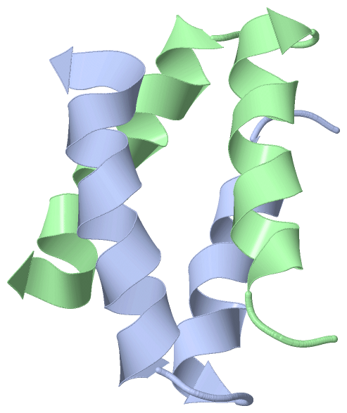
Image NMR Structure - model 1
NMR Structure - model 1  (Jmol Viewer)
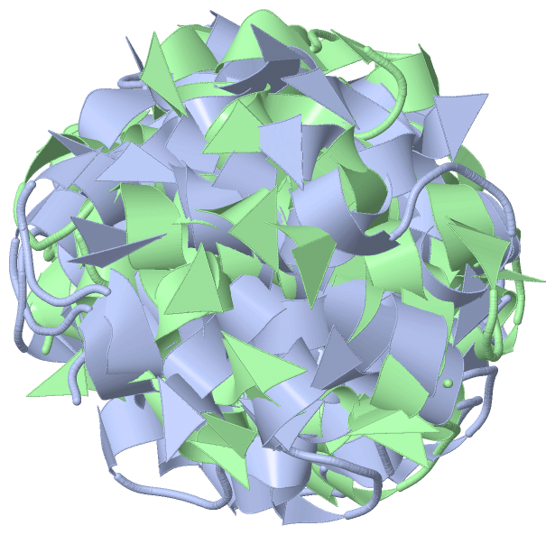
Image NMR Structure - all models
NMR Structure - all models  (Jmol Viewer)

(-) Description

Title :  SOLUTION STRUCTURE OF ALPHA2D
 
Authors :  R. B. Hill, W. F. Degrado
Date :  01 Jun 99  (Deposition) - 09 Jun 99  (Release) - 24 Feb 09  (Revision)
Method :  SOLUTION NMR
Resolution :  NOT APPLICABLE
Chains :  NMR Structure  :  A,B  (16x)
Keywords :  De Novo Design, Protein Design, Protein Folding, Bisecting U Motif, Four-Helix Bundle, Helix-Turn-Helix, De Novo Protein (Keyword Search: [Gene Ontology, PubMed, Web (Google)] )
 
Reference :  R. B. Hill, W. F. Degrado
Solution Structure Of Alpha2D, A Nativelike De Novo Designed Protein
J. Am. Chem. Soc. V. 120 1138 1998
PubMed: search  |  Reference-DOI: 10.1021/JA9733649
(for further references see the PDB file header)

(-) Compounds

Molecule 1 - PROTEIN (ALPHA2D)
    Chains: A, B
    Engineered: YES
    Other Details: THE PEPTIDE WAS CHEMICALLY SYNTHESIZED. THIS SEQUENCE WAS DESIGNED TO FOLD INTO A HELIX-TURN-HELIX PEPTIDE THAT DIMERIZES TO FORM A FOUR-HELIX BUNDLE.
    Synthetic: YES

 Structural Features

(-) Chains, Units

  
NMR Structure (16x): 

Summary Information (see also Sequences/Alignments below)

(-) Ligands, Modified Residues, Ions  (0, 0)

(no "Ligand,Modified Residues,Ions" information available for 1QP6)

(-) Sites  (0, 0)

(no "Site" information available for 1QP6)

(-) SS Bonds  (0, 0)

(no "SS Bond" information available for 1QP6)

(-) Cis Peptide Bonds  (0, 0)

(no "Cis Peptide Bond" information available for 1QP6)

 Sequence-Structure Mapping

(-) SAPs(SNPs)/Variants  (0, 0)

(no "SAP(SNP)/Variant" information available for 1QP6)

(-) PROSITE Motifs  (0, 0)

(no "PROSITE Motif" information available for 1QP6)

(-) Exons   (0, 0)

(no "Exon" information available for 1QP6)

(-) Sequences/Alignments

NMR Structure
   Reformat: Number of residues per line =  ('0' or empty: single-line sequence representation)
  Number of residues per labelling interval =   
  UniProt sequence: complete  aligned part    
   Show mapping: SCOP domains CATH domains Pfam domains Secondary structure (by author)
SAPs(SNPs) PROSITE motifs Exons
(details for a mapped element are shown in a popup box when the mouse pointer rests over it)
Chain A from PDB  Type:PROTEIN  Length:35
                                                                  
               SCOP domains d1qp6a_ A: alpha2d                  SCOP domains
               CATH domains ----------------------------------- CATH domains
               Pfam domains ----------------------------------- Pfam domains
         Sec.struct. author ..hhhhhhhhhhhhh...hhhhhhhhhhhhhhhh. Sec.struct. author
                 SAPs(SNPs) ----------------------------------- SAPs(SNPs)
                    PROSITE ----------------------------------- PROSITE
                 Transcript ----------------------------------- Transcript
                  1qp6 A  1 GEVEELEKKFKELWKGPRRGEIEELHKKFHELIKG 35
                                    10        20        30     

Chain B from PDB  Type:PROTEIN  Length:35
                                                                  
               SCOP domains d1qp6b_ B: alpha2d                  SCOP domains
               CATH domains ----------------------------------- CATH domains
               Pfam domains ----------------------------------- Pfam domains
         Sec.struct. author ..hhhhhhhhhhhhh...hhhhhhhhhhhhhhhh. Sec.struct. author
                 SAPs(SNPs) ----------------------------------- SAPs(SNPs)
                    PROSITE ----------------------------------- PROSITE
                 Transcript ----------------------------------- Transcript
                  1qp6 B  1 GEVEELEKKFKELWKGPRRGEIEELHKKFHELIKG 35
                                    10        20        30     

   Legend:   → Mismatch (orange background)
  - → Gap (green background, '-', border residues have a numbering label)
    → Modified Residue (blue background, lower-case, 'x' indicates undefined single-letter code, labelled with number + name)
  x → Chemical Group (purple background, 'x', labelled with number + name, e.g. ACE or NH2)
  extra numbering lines below/above indicate numbering irregularities and modified residue names etc., number ends below/above '|'

 Classification and Annotation

(-) SCOP Domains  (1, 2)

NMR Structure

(-) CATH Domains  (0, 0)

(no "CATH Domain" information available for 1QP6)

(-) Pfam Domains  (0, 0)

(no "Pfam Domain" information available for 1QP6)

(-) Gene Ontology  (0, 0)

NMR Structure(hide GO term definitions)
    (no "Gene Ontology" information available for 1QP6)

 Visualization

(-) Interactive Views

NMR Structure
  Complete Structure
    Jena3D(integrated viewing of ligand, site, SAP, PROSITE, SCOP information)
    WebMol | AstexViewer[tm]@PDBe
(Java Applets, require no local installation except for Java; loading may be slow)
    STRAP
(Java WebStart application, automatic local installation, requires Java; full application with system access!)
    RasMol
(require local installation)
    Molscript (VRML)
(requires installation of a VRML viewer; select preferred view via VRML and generate a mono or stereo PDF format file)
 
  Ligands, Modified Residues, Ions
(no "Ligands, Modified Residues, Ions" information available for 1qp6)
 
  Sites
(no "Sites" information available for 1qp6)
 
  Cis Peptide Bonds
(no "Cis Peptide Bonds" information available for 1qp6)
 

(-) Still Images

Jmol
  protein: cartoon or spacefill or dots and stick; nucleic acid: cartoon and stick; ligands: spacefill; active site: stick
Molscript
  protein, nucleic acid: cartoon; ligands: spacefill; active site: ball and stick

 Databases and Analysis Tools

(-) Databases

Access by PDB/NDB ID
  1qp6
    Family and Domain Information: ProDom | SYSTERS
    General Structural Information: GlycoscienceDB | MMDB | NDB | OCA | PDB | PDBe | PDBj | PDBsum | PDBWiki | PQS | PROTEOPEDIA
    Orientation in Membranes: OPM
    Protein Surface: SURFACE
    Secondary Structure: DSSP (structure derived) | HSSP (homology derived)
    Structural Genomics: GeneCensus
    Structural Neighbours: CE | VAST
    Structure Classification: CATH | Dali | SCOP
    Validation and Original Data: BMRB Data View | BMRB Restraints Grid | EDS | PROCHECK | RECOORD | WHAT_CHECK
 
Access by UniProt ID/Accession number
  (no 'UniProt ID/Accession number' available) |
    Comparative Protein Structure Models: ModBase
    Genomic Information: Ensembl
    Protein-protein Interaction: DIP
    Sequence, Family and Domain Information: InterPro | Pfam | SMART | UniProt
 
Access by Enzyme Classificator   (EC Number)
  (no 'Enzyme Classificator' available)
    General Enzyme Information: BRENDA | EC-PDB | Enzyme | IntEnz
    Pathway: KEGG | MetaCyc
 
Access by Disease Identifier   (MIM ID)
  (no 'MIM ID' available)
    Disease Information: OMIM
 
Access by GenAge ID
  (no 'GenAge ID' available)
    Age Related Information: GenAge

(-) Analysis Tools

Access by PDB/NDB ID
    Domain Information: XDom
    Interatomic Contacts of Structural Units: CSU
    Ligand-protein Contacts: LPC
    Protein Cavities: castP
    Sequence and Secondary Structure: PDBCartoon
    Structure Alignment: STRAP(Java WebStart application, automatic local installation, requires Java; full application with system access!)
    Structure and Sequence Browser: STING
 
Access by UniProt ID/Accession number
  (no 'UniProt ID/Accession number' available) |
    Protein Disorder Prediction: DisEMBL | FoldIndex | GLOBPLOT

 Related Entries

(-) Entries Sharing at Least One Protein Chain (UniProt ID)

(no "Entries Sharing at Least One Protein Chain" available for 1QP6)

(-) Related Entries Specified in the PDB File

(no "Related Entries Specified in the PDB File" available for 1QP6)