| molecular function |
|---|
| | GO:0005524 | | ATP binding | | Interacting selectively and non-covalently with ATP, adenosine 5'-triphosphate, a universally important coenzyme and enzyme regulator. |
| | GO:0003824 | | catalytic activity | | Catalysis of a biochemical reaction at physiological temperatures. In biologically catalyzed reactions, the reactants are known as substrates, and the catalysts are naturally occurring macromolecular substances known as enzymes. Enzymes possess specific binding sites for substrates, and are usually composed wholly or largely of protein, but RNA that has catalytic activity (ribozyme) is often also regarded as enzymatic. |
| | GO:0000166 | | nucleotide binding | | Interacting selectively and non-covalently with a nucleotide, any compound consisting of a nucleoside that is esterified with (ortho)phosphate or an oligophosphate at any hydroxyl group on the ribose or deoxyribose. |
| | GO:0016491 | | oxidoreductase activity | | Catalysis of an oxidation-reduction (redox) reaction, a reversible chemical reaction in which the oxidation state of an atom or atoms within a molecule is altered. One substrate acts as a hydrogen or electron donor and becomes oxidized, while the other acts as hydrogen or electron acceptor and becomes reduced. |
| | GO:0004748 | | ribonucleoside-diphosphate reductase activity, thioredoxin disulfide as acceptor | | Catalysis of the reaction: 2'-deoxyribonucleoside diphosphate + thioredoxin disulfide + H2O = ribonucleoside diphosphate + thioredoxin. Thioredoxin disulfide is the oxidized form of thioredoxin. |
| biological process |
|---|
| | GO:0006260 | | DNA replication | | The cellular metabolic process in which a cell duplicates one or more molecules of DNA. DNA replication begins when specific sequences, known as origins of replication, are recognized and bound by initiation proteins, and ends when the original DNA molecule has been completely duplicated and the copies topologically separated. The unit of replication usually corresponds to the genome of the cell, an organelle, or a virus. The template for replication can either be an existing DNA molecule or RNA. |
| | GO:0009263 | | deoxyribonucleotide biosynthetic process | | The chemical reactions and pathways resulting in the formation of a deoxyribonucleotide, a compound consisting of deoxyribonucleoside (a base linked to a deoxyribose sugar) esterified with a phosphate group at either the 3' or 5'-hydroxyl group of the sugar. |
| | GO:0008152 | | metabolic process | | The chemical reactions and pathways, including anabolism and catabolism, by which living organisms transform chemical substances. Metabolic processes typically transform small molecules, but also include macromolecular processes such as DNA repair and replication, and protein synthesis and degradation. |
| | GO:0015949 | | nucleobase-containing small molecule interconversion | | The chemical reactions and pathways by which a nucleobase, nucleoside or nucleotide small molecule is synthesized from another nucleobase, nucleoside or nucleotide small molecule. |
| | GO:0055114 | | oxidation-reduction process | | A metabolic process that results in the removal or addition of one or more electrons to or from a substance, with or without the concomitant removal or addition of a proton or protons. |
| cellular component |
|---|
| | GO:0005829 | | cytosol | | The part of the cytoplasm that does not contain organelles but which does contain other particulate matter, such as protein complexes. |
| | GO:0005971 | | ribonucleoside-diphosphate reductase complex | | An enzyme complex composed of 2-4 or more subunits, which usually contains nonheme iron and requires ATP for catalysis. Catalyzes the formation of 2'-deoxyribonucleoside diphosphate from ribonucleoside diphosphate, using either thioredoxin disulfide or glutaredoxin disulfide as an acceptor. |
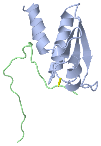
 Description
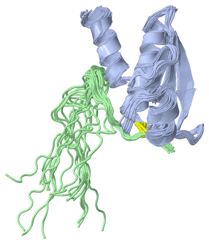