Show PDB file:   
         Plain Text   HTML   (compressed file size)
QuickSearch:   
by PDB,NDB,UniProt,PROSITE Code or Search Term(s)  
(-)Asym./Biol. Unit
(-)Asym./Biol. Unit - sites
collapse expand < >
Image Asym./Biol. Unit
Asym./Biol. Unit  (Jmol Viewer)
Image Asym./Biol. Unit - sites
Asym./Biol. Unit - sites  (Jmol Viewer)

(-) Description

Title :  CRYSTAL STRUCTURE OF PASTEURELLA HAEMOLYTICA APO FERRIC ION-BINDING PROTEIN A
 
Authors :  S. R. Shouldice, D. R. Dougan, R. J. Skene, G. Snell, D. Scheibe, P. A. Williams, S. Kirby, D. E. Mcree, A. B. Schryvers, L. W. Tari
Date :  28 Jul 03  (Deposition) - 11 Nov 03  (Release) - 24 Feb 09  (Revision)
Method :  X-RAY DIFFRACTION
Resolution :  1.20
Chains :  Asym./Biol. Unit :  A
Keywords :  Iron Binding Protein, Metal Binding Protein (Keyword Search: [Gene Ontology, PubMed, Web (Google))
 
Reference :  S. R. Shouldice, D. R. Dougan, P. A. Williams, R. J. Skene, G. Snell, D. Scheibe, S. Kirby, D. J. Hosfield, D. E. Mcree, A. B. Schryvers, L. W. Tari
Crystal Structure Of Pasteurella Haemolytica Ferric Ion-Binding Protein A Reveals A Novel Class Of Bacterial Iron-Binding Proteins
J. Biol. Chem. V. 278 41093 2003
PubMed-ID: 12882966  |  Reference-DOI: 10.1074/JBC.M306821200
(for further references see the PDB file header)

(-) Compounds

Molecule 1 - IRON BINDING PROTEIN FBPA
    ChainsA
    EngineeredYES
    Expression SystemESCHERICHIA COLI
    Expression System PlasmidPT7-7
    Expression System StrainBL21(DE3)/PLYSS
    Expression System Taxid562
    Expression System Vector TypePLASMID
    GeneFBPA
    Organism ScientificMANNHEIMIA HAEMOLYTICA
    Organism Taxid75985
    SynonymFERRIC ION-BINDING PROTEIN A

 Structural Features

(-) Chains, Units

  1
Asymmetric/Biological Unit A

Summary Information (see also Sequences/Alignments below)
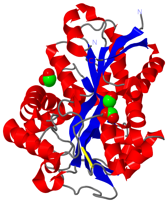
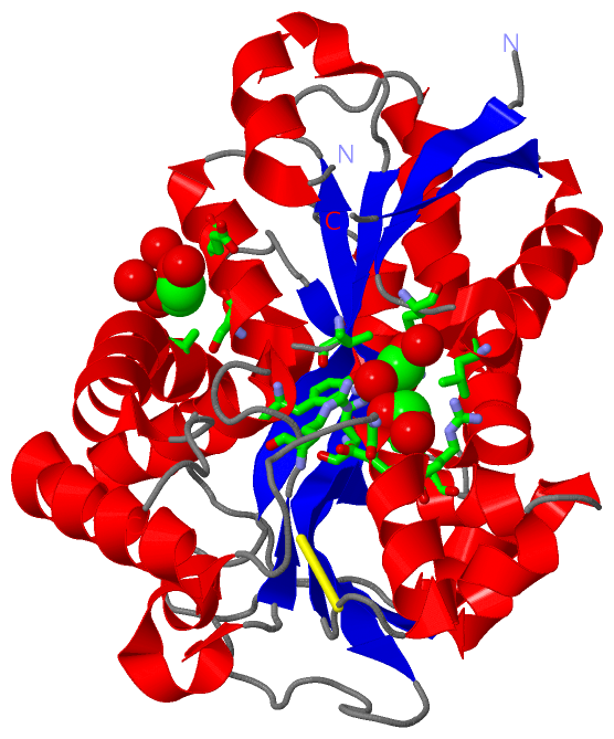
(-) Ligands, Modified Residues, Ions  (2, 3)

Asymmetric/Biological Unit (2, 3)
No.NameCountTypeFull Name
1EDO1Ligand/Ion1,2-ETHANEDIOL
2FMT2Ligand/IonFORMIC ACID

(-) Sites  (3, 3)

Asymmetric Unit (3, 3)
No.NameEvidenceResiduesDescription
1AC1SOFTWARESER A:61 , GLU A:65 , ALA A:298 , HOH A:1198 , HOH A:1261 , HOH A:1337 , HOH A:1400BINDING SITE FOR RESIDUE EDO A 1003
2AC2SOFTWAREARG A:10 , LEU A:14 , VAL A:58 , ARG A:101 , TYR A:142 , TYR A:198 , HOH A:1164BINDING SITE FOR RESIDUE FMT A 1001
3AC3SOFTWAREARG A:136 , GLY A:177 , ASP A:178 , ARG A:179 , TYR A:199 , HOH A:1020 , HOH A:1172BINDING SITE FOR RESIDUE FMT A 1002

(-) SS Bonds  (1, 1)

Asymmetric/Biological Unit
No.Residues
1A:134 -A:190

(-) Cis Peptide Bonds  (1, 1)

Asymmetric/Biological Unit
No.Residues
1Gly A:38 -Leu A:39

 Sequence-Structure Mapping

(-) SAPs(SNPs)/Variants  (0, 0)

(no "SAP(SNP)/Variant" information available for 1Q35)

(-) PROSITE Motifs  (0, 0)

(no "PROSITE Motif" information available for 1Q35)

(-) Exons   (0, 0)

(no "Exon" information available for 1Q35)

(-) Sequences/Alignments

Asymmetric/Biological Unit
   Reformat: Number of residues per line =  ('0' or empty: single-line sequence representation)
  Number of residues per labelling interval =   
  UniProt sequence: complete  aligned part    
   Show mapping: SCOP domains CATH domains Pfam domains Secondary structure (by author)
SAPs(SNPs) PROSITE motifs Exons
(details for a mapped element are shown in a popup box when the mouse pointer rests over it)
Chain A from PDB  Type:PROTEIN  Length:317
 aligned with Q9Z4N6_MANHA | Q9Z4N6 from UniProtKB/TrEMBL  Length:342

    Alignment length:318
                                    32        42        52        62        72        82        92       102       112       122       132       142       152       162       172       182       192       202       212       222       232       242       252       262       272       282       292       302       312       322       332        
         Q9Z4N6_MANHA    23 ANEVNVYSYRQPYLIEPMLKNFEKDTGIKVNIIFADKGLVDRVKQEGELSPADVLLTVDISRVMEIVNADLAQKIDSKVLEKNIPAQFRDSNDQWFGLTTRARVIYTSKDRVGKLPAGFDYLDLAKPEYKGKVCVRSGKNSYNVSLFAAMIEHYGIEKTKAFLEGLKANLARKPQGGDRDQVKAIKEGICDYSIGNSYYYGKMLDDEKQKSWAEAAIINFPSGEHGTHKNISGVVIAKHSPNKANAVKLIEYLSGEKAQGLYAELNHEYPVKEGIEPSAIVKGWGTFKSDTIKLEDIAKNYEAALKLVDEVKFDDFSE 340
               SCOP domains d1q35a_ A: Ferric-binding protein Fb pA                                                                                                                                                                                                                                                                                        SCOP domains
               CATH domains 1q35A01 A:1-98,A:227-275 Periplasmic  binding protein-like II                                     1q35A02 A:99-226,A:276-318 Periplasmic binding protein-like II                                                                  1q35A01 A:1-98,A:227-275                         1q35A02 A:99-226,A:276-318                  CATH domains
               Pfam domains ------------------------------------------------------------------------------------------------------------------------------------------------------------------------------------------------------------------------------------------------------------------------------------------------------------------------------ Pfam domains
         Sec.struct. author ..eeeeee..hhhhhhhhhhhhhhhhh.eeeeee..-.hhhhhhhhhhhh...eeee.hhhhhhhhhhh.......hhhhhhhhhhhhh......eeeeeeeeeeeee...........hhhhhhhhhhh..ee.....hhhhhhhhhhhhhhhhhhhhhhhhhhhhhh.......hhhhhhhhhhh....eeeeehhhhhhhhhh..hhhhhhheeee........eeeeeeeee.....hhhhhhhhhhhhhhhhhhhhhhhhhh.ee.......hhhhhhh........hhhhhhhhhhhhhhhhhhhh...... Sec.struct. author
                 SAPs(SNPs) ------------------------------------------------------------------------------------------------------------------------------------------------------------------------------------------------------------------------------------------------------------------------------------------------------------------------------ SAPs(SNPs)
                    PROSITE ------------------------------------------------------------------------------------------------------------------------------------------------------------------------------------------------------------------------------------------------------------------------------------------------------------------------------ PROSITE
                 Transcript ------------------------------------------------------------------------------------------------------------------------------------------------------------------------------------------------------------------------------------------------------------------------------------------------------------------------------ Transcript
                 1q35 A   1 ANEVNVYSYRQPYLIEPMLKNFEKDTGIKVNIIFAD-GLVDRVKQEGELSPADVLLTVDISRVMEIVNADLAQKIDSKVLEKNIPAQFRDSNDQWFGLTTRARVIYTSKDRVGKLPAGFDYLDLAKPEYKGKVCVRSGKNSYNVSLFAAMIEHYGIEKTKAFLEGLKANLARKPQGGDRDQVKAIKEGICDYSIGNSYYYGKMLDDEKQKSWAEAAIINFPSGEHGTHKNISGVVIAKHSPNKANAVKLIEYLSGEKAQGLYAELNHEYPVKEGIEPSAIVKGWGTFKSDTIKLEDIAKNYEAALKLVDEVKFDDFSE 318
                                    10        20        30     | |40        50        60        70        80        90       100       110       120       130       140       150       160       170       180       190       200       210       220       230       240       250       260       270       280       290       300       310        
                                                              36 |                                                                                                                                                                                                                                                                                        
                                                                38                                                                                                                                                                                                                                                                                        

   Legend:   → Mismatch (orange background)
  - → Gap (green background, '-', border residues have a numbering label)
    → Modified Residue (blue background, lower-case, 'x' indicates undefined single-letter code, labelled with number + name)
  x → Chemical Group (purple background, 'x', labelled with number + name, e.g. ACE or NH2)
  extra numbering lines below/above indicate numbering irregularities and modified residue names etc., number ends below/above '|'

 Classification and Annotation

(-) SCOP Domains  (1, 1)

Asymmetric/Biological Unit

(-) CATH Domains  (1, 2)

Asymmetric/Biological Unit
(-)
Class: Alpha Beta (26913)

(-) Pfam Domains  (0, 0)

(no "Pfam Domain" information available for 1Q35)

(-) Gene Ontology  (1, 1)

Asymmetric/Biological Unit(hide GO term definitions)
Chain A   (Q9Z4N6_MANHA | Q9Z4N6)
molecular function
    GO:0046872    metal ion binding    Interacting selectively and non-covalently with any metal ion.

 Visualization

(-) Interactive Views

Asymmetric/Biological Unit
  Complete Structure
    Jena3D(integrated viewing of ligand, site, SAP, PROSITE, SCOP information)
    WebMol | AstexViewer[tm]@PDBe
(Java Applets, require no local installation except for Java; loading may be slow)
    STRAP
(Java WebStart application, automatic local installation, requires Java; full application with system access!)
    RasMol
(require local installation)
    Molscript (VRML)
(requires installation of a VRML viewer; select preferred view via VRML and generate a mono or stereo PDF format file)
 
  Ligands, Modified Residues, Ions
    EDO  [ RasMol | Jena3D ]  +environment [ RasMol | Jena3D ]
    FMT  [ RasMol | Jena3D ]  +environment [ RasMol | Jena3D ]
 
  Sites
    AC1  [ RasMol ]  +environment [ RasMol ]
    AC2  [ RasMol ]  +environment [ RasMol ]
    AC3  [ RasMol ]  +environment [ RasMol ]
 
  Cis Peptide Bonds
    Gly A:38 - Leu A:39   [ RasMol ]  
 

(-) Still Images

Jmol
  protein: cartoon or spacefill or dots and stick; nucleic acid: cartoon and stick; ligands: spacefill; active site: stick
Molscript
  protein, nucleic acid: cartoon; ligands: spacefill; active site: ball and stick

 Databases and Analysis Tools

(-) Databases

Access by PDB/NDB ID
  1q35
    Family and Domain InformationProDom | SYSTERS
    General Structural InformationGlycoscienceDB | MMDB | NDB | OCA | PDB | PDBe | PDBj | PDBsum | PDBWiki | PQS | PROTEOPEDIA
    Orientation in MembranesOPM
    Protein SurfaceSURFACE
    Secondary StructureDSSP (structure derived) | HSSP (homology derived)
    Structural GenomicsGeneCensus
    Structural NeighboursCE | VAST
    Structure ClassificationCATH | Dali | SCOP
    Validation and Original DataBMRB Data View | BMRB Restraints Grid | EDS | PROCHECK | RECOORD | WHAT_CHECK
 
Access by UniProt ID/Accession number
  Q9Z4N6_MANHA | Q9Z4N6
    Comparative Protein Structure ModelsModBase
    Genomic InformationEnsembl
    Protein-protein InteractionDIP
    Sequence, Family and Domain InformationInterPro | Pfam | SMART | UniProtKB/TrEMBL
 
Access by Enzyme Classificator   (EC Number)
  (no 'Enzyme Classificator' available)
    General Enzyme InformationBRENDA | EC-PDB | Enzyme | IntEnz
    PathwayKEGG | MetaCyc
 
Access by Disease Identifier   (MIM ID)
  (no 'MIM ID' available)
    Disease InformationOMIM
 
Access by GenAge ID
  (no 'GenAge ID' available)
    Age Related InformationGenAge

(-) Analysis Tools

Access by PDB/NDB ID
    Domain InformationXDom
    Interatomic Contacts of Structural UnitsCSU
    Ligand-protein ContactsLPC
    Protein CavitiescastP
    Sequence and Secondary StructurePDBCartoon
    Structure AlignmentSTRAP(Java WebStart application, automatic local installation, requires Java; full application with system access!)
    Structure and Sequence BrowserSTING
 
Access by UniProt ID/Accession number
  Q9Z4N6_MANHA | Q9Z4N6
    Protein Disorder PredictionDisEMBL | FoldIndex | GLOBPLOT (for more information see DisProt)

 Related Entries

(-) Entries Sharing at Least One Protein Chain (UniProt ID)

UniProtKB/TrEMBL
        Q9Z4N6_MANHA | Q9Z4N61si0 1si1

(-) Related Entries Specified in the PDB File

(no "Related Entries Specified in the PDB File" available for 1Q35)