|
|
|
|
 Description
|
|
Compounds
|
||||||||||||||||||||||||||||||||||||||||
Chains, Units
Summary Information (see also Sequences/Alignments below) |
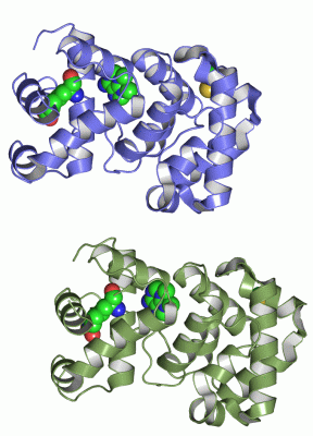
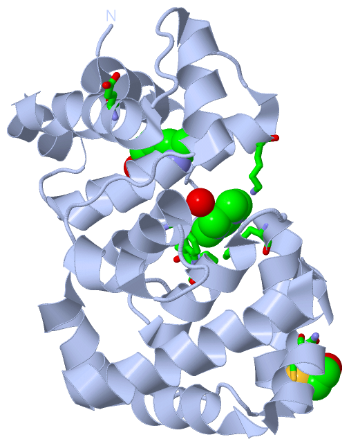
Ligands, Modified Residues, Ions (3, 6)
Asymmetric Unit (3, 6)
|
Sites (4, 4)
Asymmetric Unit (4, 4)
|
SS Bonds (0, 0)| (no "SS Bond" information available for 1PU7) |
Cis Peptide Bonds (0, 0)| (no "Cis Peptide Bond" information available for 1PU7) |
SAPs(SNPs)/Variants (0, 0)| (no "SAP(SNP)/Variant" information available for 1PU7) |
PROSITE Motifs (0, 0)| (no "PROSITE Motif" information available for 1PU7) |
Exons (0, 0)| (no "Exon" information available for 1PU7) |
Sequences/Alignments
Asymmetric UnitChain A from PDB Type:PROTEIN Length:215 aligned with O25323_HELPY | O25323 from UniProtKB/TrEMBL Length:218 Alignment length:215 11 21 31 41 51 61 71 81 91 101 111 121 131 141 151 161 171 181 191 201 211 O25323_HELPY 2 LDSFEILKALKSLDLLKNAPSWWWPNALKFEALLGAVLTQNTKFEAVLKSLENLKNAFILENDDEINLKKIAYIEFSKLAECVRPSGFYNQKAKRLIDLSKNILKDFQSFENFKQEVTREWLLNQKGVGKESADAILCYVCAKEVMVVDKYSYLFLKKIGIEIEDYDELQHFFEKGVQENLNSALALYENTIPLAQLYARFHGKIVEFSKQKLEL 216 SCOP domains d1pu7a_ A: 3-Methyladenine DNA glycosylase III (MagIII) SCOP domains CATH domains 1pu7A01 1pu7A02 A:22-144 Hypothetical protein; domain 2 1pu7A01 A:2-21,A:145-216 CATH domains Pfam domains ----------------------------------------------------------------------------------------------------------------------------------------------------------------------------------------------------------------------- Pfam domains SAPs(SNPs) ----------------------------------------------------------------------------------------------------------------------------------------------------------------------------------------------------------------------- SAPs(SNPs) PROSITE ----------------------------------------------------------------------------------------------------------------------------------------------------------------------------------------------------------------------- PROSITE Transcript ----------------------------------------------------------------------------------------------------------------------------------------------------------------------------------------------------------------------- Transcript 1pu7 A 2 LDSFEILKALKSLDLLKNAPAWWWPNALKFEALLGAVLTQNTKFEAVLKSLENLKNAFILENDDEINLKKIAYIEFSKLAECVRPSGFYNQKAKRLIDLSGNILKDFQSFENFKQEVTREWLLDQKGIGKESADAILCYACAKEVMVVDKYSYLFLKKLGIEIEDYDELQHFFEKGVQENLNSALALYENTISLAQLYARFHGkIVEFSKQKLEL 216 11 21 31 41 51 61 71 81 91 101 111 121 131 141 151 161 171 181 191 201 | 211 205-KCX Chain B from PDB Type:PROTEIN Length:216 aligned with O25323_HELPY | O25323 from UniProtKB/TrEMBL Length:218 Alignment length:216 10 20 30 40 50 60 70 80 90 100 110 120 130 140 150 160 170 180 190 200 210 O25323_HELPY 1 MLDSFEILKALKSLDLLKNAPSWWWPNALKFEALLGAVLTQNTKFEAVLKSLENLKNAFILENDDEINLKKIAYIEFSKLAECVRPSGFYNQKAKRLIDLSKNILKDFQSFENFKQEVTREWLLNQKGVGKESADAILCYVCAKEVMVVDKYSYLFLKKIGIEIEDYDELQHFFEKGVQENLNSALALYENTIPLAQLYARFHGKIVEFSKQKLEL 216 SCOP domains d1pu7b_ B: 3-Methyladenine DNA glycosylase III (MagIII) SCOP domains CATH domains 1pu7B01 1pu7B02 B:22-144 Hypothetical protein; domain 2 1pu7B01 B:1-21,B:145-216 CATH domains Pfam domains (1) ----------------------------------HhH-GPD-1pu7B01 B:35-180 ------------------------------------ Pfam domains (1) Pfam domains (2) ----------------------------------HhH-GPD-1pu7B02 B:35-180 ------------------------------------ Pfam domains (2) SAPs(SNPs) ------------------------------------------------------------------------------------------------------------------------------------------------------------------------------------------------------------------------ SAPs(SNPs) PROSITE ------------------------------------------------------------------------------------------------------------------------------------------------------------------------------------------------------------------------ PROSITE Transcript ------------------------------------------------------------------------------------------------------------------------------------------------------------------------------------------------------------------------ Transcript 1pu7 B 1 VLDSFEILKALKSLDLLKNAPAWWWPNALKFEALLGAVLTQNTKFEAVLKSLENLKNAFILENDDEINLKKIAYIEFSKLAECVRPSGFYNQKAKRLIDLSGNILKDFQSFENFKQEVTREWLLDQKGIGKESADAILCYACAKEVMVVDKYSYLFLKKLGIEIEDYDELQHFFEKGVQENLNSALALYENTISLAQLYARFHGkIVEFSKQKLEL 216 10 20 30 40 50 60 70 80 90 100 110 120 130 140 150 160 170 180 190 200 | 210 205-KCX
|
||||||||||||||||||||
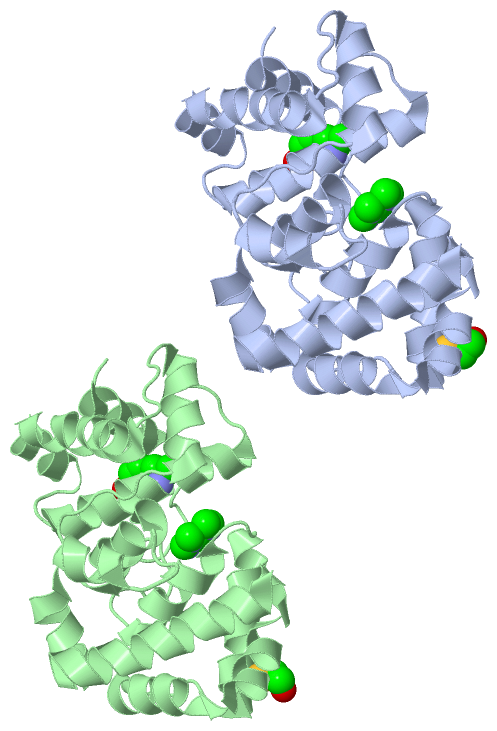
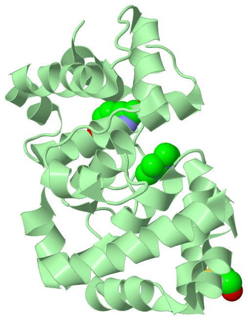
SCOP Domains (1, 2)
Asymmetric Unit
 |
CATH Domains (2, 4)
Asymmetric Unit
|
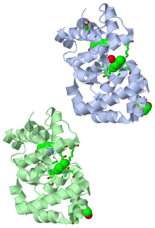
Pfam Domains (1, 2)
Asymmetric Unit
|
Gene Ontology (6, 6)|
Asymmetric Unit(hide GO term definitions) Chain A,B (O25323_HELPY | O25323)
|
||||||||||||||||||||||||||||||||||||||||||||||||
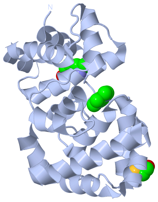
Interactive Views
|
||||||||||||||||||||||||||||||||||||||||||||||||||||||||||||||||||||||||||||||||||||||||||||||||||||||||||||||||||||||||||||||||||||||||||||||||||||||||||||||||||||||||||||||||
Still Images
|
||||||||||||||||
Databases
|
||||||||||||||||||||||||||||||||||||||||||||||||||||||||||||||||||||||||||||||||||||||||||||||||||||||||||||||||||||||||||||||||||||||||||||||||||||||||||||||||
Analysis Tools
|
|||||||||||||||||||||||||||||||||||||||||||||||||||||||||||||
Entries Sharing at Least One Protein Chain (UniProt ID)
Related Entries Specified in the PDB File
|
|