Show PDB file:   
         Plain Text   HTML   (compressed file size)
QuickSearch:   
by PDB,NDB,UniProt,PROSITE Code or Search Term(s)  
(-)NMR Structure - model 1
(-)NMR Structure - model 1, sites
(-)NMR Structure - all models
collapse expand < >
Image NMR Structure - model 1
NMR Structure - model 1  (Jmol Viewer)
Image NMR Structure - model 1, sites
NMR Structure - model 1, sites  (Jmol Viewer)
Image NMR Structure - all models
NMR Structure - all models  (Jmol Viewer)

(-) Description

Title :  SOLUTION CONFORMATION OF ALPHAA-CONOTOXIN EIVA
 
Authors :  S. -W. Chi, K. -H. Park, J. -E. Suk, B. M. Olivera, J. M. Mcintosh, K. - H. Han
Date :  18 Jun 03  (Deposition) - 04 Nov 03  (Release) - 24 Feb 09  (Revision)
Method :  SOLUTION NMR
Resolution :  NOT APPLICABLE
Chains :  NMR Structure  :  A  (18x)
Keywords :  Alpha-Helix, Two Disulfide Bonds, C-Term Amidation, Toxin (Keyword Search: [Gene Ontology, PubMed, Web (Google))
 
Reference :  S. -W. Chi, K. -H. Park, J. -E. Suk, B. M. Olivera, J. M. Mcintosh, K. -H. Han
Solution Conformation Of Alphaa-Conotoxin Eiva, A Potent Neuromuscular Nicotinic Acetylcholine Receptor Antagonist From Conus Ermineus
J. Biol. Chem. V. 278 42208 2003
PubMed-ID: 12900418  |  Reference-DOI: 10.1074/JBC.M303342200
(for further references see the PDB file header)

(-) Compounds

Molecule 1 - ALPHA-A-CONOTOXIN EIVA
    ChainsA
    EngineeredYES
    Other DetailsTHIS SEQUENCE OCCURS NATURALLY IN CONUS ERMINEUS VENOM.
    SyntheticYES

 Structural Features

(-) Chains, Units

  
NMR Structure (18x)

Summary Information (see also Sequences/Alignments below)

(-) Ligands, Modified Residues, Ions  (2, 6)

NMR Structure (2, 6)
No.NameCountTypeFull Name
1HYP5Mod. Amino Acid4-HYDROXYPROLINE
2NH21Mod. Amino AcidAMINO GROUP

(-) Sites  (1, 1)

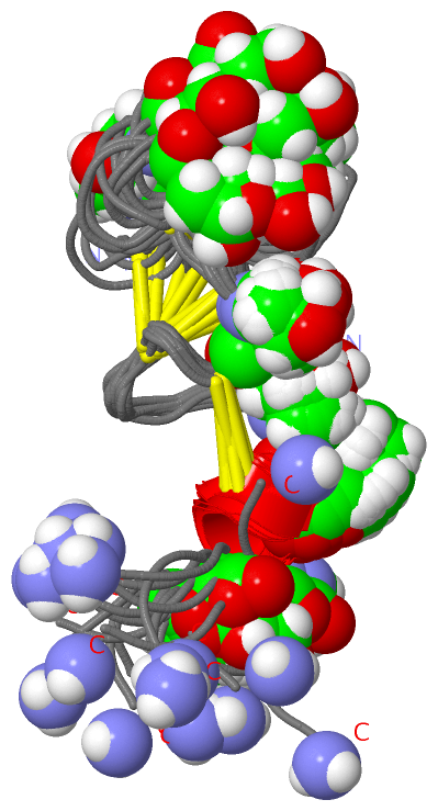
NMR Structure (1, 1)
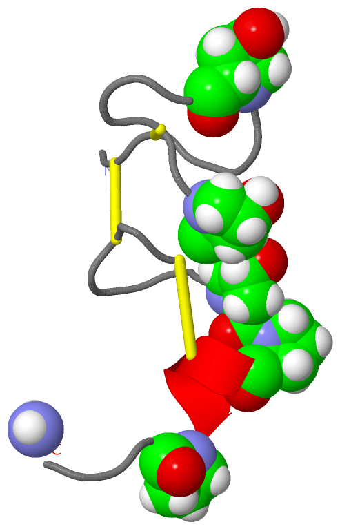
No.NameEvidenceResiduesDescription
1AC1SOFTWAREGLY A:30BINDING SITE FOR RESIDUE NH2 A 31

(-) SS Bonds  (3, 3)

NMR Structure
No.Residues
1A:2 -A:16
2A:3 -A:11
3A:14 -A:24

(-) Cis Peptide Bonds  (1, 1)

NMR Structure
No.Residues
1Gly A:1 -Cys A:2

 Sequence-Structure Mapping

(-) SAPs(SNPs)/Variants  (0, 0)

(no "SAP(SNP)/Variant" information available for 1PQR)

(-) PROSITE Motifs  (0, 0)

(no "PROSITE Motif" information available for 1PQR)

(-) Exons   (0, 0)

(no "Exon" information available for 1PQR)

(-) Sequences/Alignments

NMR Structure
   Reformat: Number of residues per line =  ('0' or empty: single-line sequence representation)
  Number of residues per labelling interval =   
  UniProt sequence: complete  aligned part    
   Show mapping: SCOP domains CATH domains Pfam domains Secondary structure (by author)
SAPs(SNPs) PROSITE motifs Exons
(details for a mapped element are shown in a popup box when the mouse pointer rests over it)
Chain A from PDB  Type:PROTEIN  Length:31
 aligned with CA4A_CONER | P58782 from UniProtKB/Swiss-Prot  Length:30

    Alignment length:31
                                                        30 
                                    10        20        30 
            CA4A_CONER    1 GCCGPYPNAACHPCGCKVGRPPYCDRPSGG-  -
               SCOP domains d1pqra_ A:                      SCOP domains
               CATH domains ------------------------------- CATH domains
               Pfam domains Toxin_14-1pqrA01 A:1-26   ----- Pfam domains
         Sec.struct. author ....................hhhhhh..... Sec.struct. author
                 SAPs(SNPs) ------------------------------- SAPs(SNPs)
                    PROSITE ------------------------------- PROSITE
                 Transcript ------------------------------- Transcript
                  1pqr A  1 GCCGPYpNAACHpCGCKVGRppYCDRpSGGx 31
                                  | 10  |     20||    | 30|
                                  7-HYP |      21-HYP |   |
                                       13-HYP   22-HYP|   |
                                                     27-HYP
                                                         31-NH2

   Legend:   → Mismatch (orange background)
  - → Gap (green background, '-', border residues have a numbering label)
    → Modified Residue (blue background, lower-case, 'x' indicates undefined single-letter code, labelled with number + name)
  x → Chemical Group (purple background, 'x', labelled with number + name, e.g. ACE or NH2)
  extra numbering lines below/above indicate numbering irregularities and modified residue names etc., number ends below/above '|'

 Classification and Annotation

(-) SCOP Domains  (1, 1)

NMR Structure
(-)
Class: Peptides (792)

(-) CATH Domains  (0, 0)

(no "CATH Domain" information available for 1PQR)

(-) Pfam Domains  (1, 1)

NMR Structure

(-) Gene Ontology  (5, 5)

NMR Structure(hide GO term definitions)
Chain A   (CA4A_CONER | P58782)
molecular function
    GO:0030550    acetylcholine receptor inhibitor activity    Interacting (directly or indirectly) with acetylcholine receptors such that the proportion of receptors in the active form is decreased.
biological process
    GO:2000272    negative regulation of receptor activity    Any process that stops, prevents or reduces the frequency, rate or extent of receptor activity.
    GO:0009405    pathogenesis    The set of specific processes that generate the ability of an organism to induce an abnormal, generally detrimental state in another organism.
cellular component
    GO:0005576    extracellular region    The space external to the outermost structure of a cell. For cells without external protective or external encapsulating structures this refers to space outside of the plasma membrane. This term covers the host cell environment outside an intracellular parasite.
    GO:0035792    other organism postsynaptic membrane    A postsynaptic membrane that is part of another organism, i.e. a secondary organism with which the first organism is interacting. A postsynaptic membrane is a specialized area of membrane facing the presynaptic membrane on the tip of the nerve ending and separated from it by a minute cleft (the synaptic cleft). Neurotransmitters transmit the signal across the synaptic cleft to the postsynaptic membrane.

 Visualization

(-) Interactive Views

NMR Structure
  Complete Structure
    Jena3D(integrated viewing of ligand, site, SAP, PROSITE, SCOP information)
    WebMol | AstexViewer[tm]@PDBe
(Java Applets, require no local installation except for Java; loading may be slow)
    STRAP
(Java WebStart application, automatic local installation, requires Java; full application with system access!)
    RasMol
(require local installation)
    Molscript (VRML)
(requires installation of a VRML viewer; select preferred view via VRML and generate a mono or stereo PDF format file)
 
  Ligands, Modified Residues, Ions
    HYP  [ RasMol | Jena3D ]  +environment [ RasMol | Jena3D ]
    NH2  [ RasMol | Jena3D ]  +environment [ RasMol | Jena3D ]
 
  Sites
    AC1  [ RasMol ]  +environment [ RasMol ]
 
  Cis Peptide Bonds
    Gly A:1 - Cys A:2   [ RasMol ]  
 

(-) Still Images

Jmol
  protein: cartoon or spacefill or dots and stick; nucleic acid: cartoon and stick; ligands: spacefill; active site: stick
Molscript
  protein, nucleic acid: cartoon; ligands: spacefill; active site: ball and stick

 Databases and Analysis Tools

(-) Databases

Access by PDB/NDB ID
  1pqr
    Family and Domain InformationProDom | SYSTERS
    General Structural InformationGlycoscienceDB | MMDB | NDB | OCA | PDB | PDBe | PDBj | PDBsum | PDBWiki | PQS | PROTEOPEDIA
    Orientation in MembranesOPM
    Protein SurfaceSURFACE
    Secondary StructureDSSP (structure derived) | HSSP (homology derived)
    Structural GenomicsGeneCensus
    Structural NeighboursCE | VAST
    Structure ClassificationCATH | Dali | SCOP
    Validation and Original DataBMRB Data View | BMRB Restraints Grid | EDS | PROCHECK | RECOORD | WHAT_CHECK
 
Access by UniProt ID/Accession number
  CA4A_CONER | P58782
    Comparative Protein Structure ModelsModBase
    Genomic InformationEnsembl
    Protein-protein InteractionDIP
    Sequence, Family and Domain InformationInterPro | Pfam | SMART | UniProtKB/SwissProt
 
Access by Enzyme Classificator   (EC Number)
  (no 'Enzyme Classificator' available)
    General Enzyme InformationBRENDA | EC-PDB | Enzyme | IntEnz
    PathwayKEGG | MetaCyc
 
Access by Disease Identifier   (MIM ID)
  (no 'MIM ID' available)
    Disease InformationOMIM
 
Access by GenAge ID
  (no 'GenAge ID' available)
    Age Related InformationGenAge

(-) Analysis Tools

Access by PDB/NDB ID
    Domain InformationXDom
    Interatomic Contacts of Structural UnitsCSU
    Ligand-protein ContactsLPC
    Protein CavitiescastP
    Sequence and Secondary StructurePDBCartoon
    Structure AlignmentSTRAP(Java WebStart application, automatic local installation, requires Java; full application with system access!)
    Structure and Sequence BrowserSTING
 
Access by UniProt ID/Accession number
  CA4A_CONER | P58782
    Protein Disorder PredictionDisEMBL | FoldIndex | GLOBPLOT (for more information see DisProt)

 Related Entries

(-) Entries Sharing at Least One Protein Chain (UniProt ID)

(no "Entries Sharing at Least One Protein Chain" available for 1PQR)

(-) Related Entries Specified in the PDB File

(no "Related Entries Specified in the PDB File" available for 1PQR)