Asymmetric Unit (11, 11)
| No. | Name | Evidence | Residues | Description |
|---|
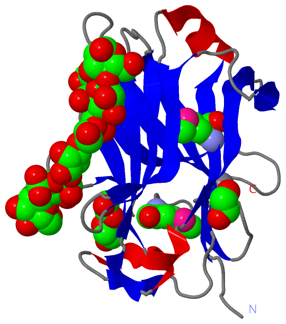
| 01 | AC1 | SOFTWARE | ASP X:111 , GLY X:112 , ARG X:155 , BMA X:187 , BMA X:190 , BMA X:191 , HOH X:409 , HOH X:434 , HOH X:442 , HOH X:486 , HOH X:679 | BINDING SITE FOR RESIDUE MAN X 186 |
| 02 | AC2 | SOFTWARE | TRP X:60 , GLU X:74 , GLN X:76 , LYS X:106 , ASP X:111 , MAN X:186 , BMA X:188 , HOH X:409 , HOH X:658 | BINDING SITE FOR RESIDUE BMA X 187 |
| 03 | AC3 | SOFTWARE | GLN X:62 , GLN X:76 , LYS X:106 , GLN X:161 , BMA X:187 , BMA X:189 , HOH X:647 , HOH X:658 | BINDING SITE FOR RESIDUE BMA X 188 |
| 04 | AC4 | SOFTWARE | TRP X:23 , TRP X:113 , LYS X:153 , BMA X:188 , BMA X:190 , HOH X:408 , HOH X:413 , HOH X:457 , HOH X:492 , HOH X:568 | BINDING SITE FOR RESIDUE BMA X 189 |
| 05 | AC5 | SOFTWARE | TRP X:23 , ASN X:110 , LYS X:153 , MAN X:186 , BMA X:189 , BMA X:191 , HOH X:434 , HOH X:529 , HOH X:682 | BINDING SITE FOR RESIDUE BMA X 190 |
| 06 | AC6 | SOFTWARE | GLU X:74 , ARG X:155 , MAN X:186 , BMA X:190 , HOH X:679 | BINDING SITE FOR RESIDUE BMA X 191 |
| 07 | AC7 | SOFTWARE | ASP X:10 , GLU X:12 , ASN X:44 , TYR X:46 , ASP X:176 , HOH X:432 | BINDING SITE FOR RESIDUE CA X 300 |
| 08 | AC8 | SOFTWARE | CYS X:29 , LYS X:31 , GLU X:53 , ASN X:55 , ASP X:61 | BINDING SITE FOR RESIDUE EDO X 401 |
| 09 | AC9 | SOFTWARE | LYS X:45 , TYR X:46 , ALA X:145 , ALA X:185 , HOH X:425 , HOH X:484 , HOH X:561 , HOH X:625 , HOH X:661 | BINDING SITE FOR RESIDUE EDO X 402 |
| 10 | BC1 | SOFTWARE | SER X:18 , LYS X:31 , TRP X:68 , GLY X:71 , VAL X:73 , EDO X:404 , HOH X:533 , HOH X:537 , HOH X:669 | BINDING SITE FOR RESIDUE EDO X 403 |
| 11 | BC2 | SOFTWARE | SER X:18 , GLU X:21 , GLY X:66 , THR X:67 , TRP X:68 , GLN X:157 , EDO X:403 , HOH X:493 | BINDING SITE FOR RESIDUE EDO X 404 |
|
 Description