|
|
|
|
 Description
|
|
Compounds
|
||||||||||||||||||||||||
Chains, Units
Summary Information (see also Sequences/Alignments below) |
Ligands, Modified Residues, Ions (1, 1)
NMR Structure (1, 1)
|
Sites (1, 1)
NMR Structure (1, 1)
|
SS Bonds (0, 0)| (no "SS Bond" information available for 1PIJ) |
Cis Peptide Bonds (0, 0)| (no "Cis Peptide Bond" information available for 1PIJ) |
SAPs(SNPs)/Variants (0, 0)| (no "SAP(SNP)/Variant" information available for 1PIJ) |
PROSITE Motifs (1, 1)
NMR Structure (1, 1)
|
||||||||||||||||||||||||
Exons (0, 0)| (no "Exon" information available for 1PIJ) |
Sequences/Alignments
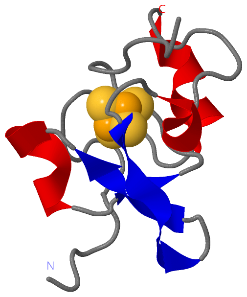
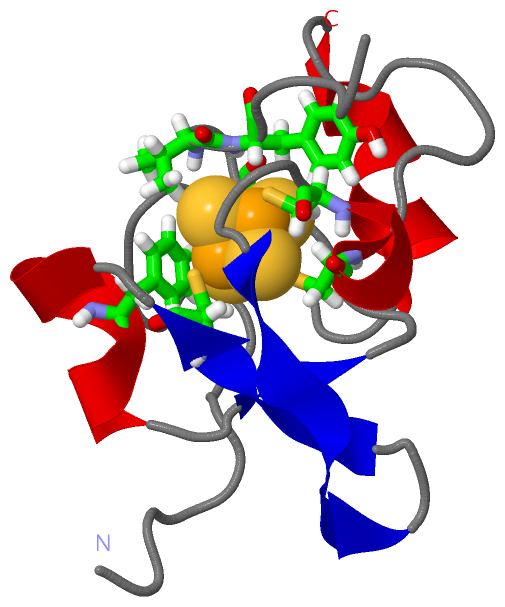
NMR StructureChain A from PDB Type:PROTEIN Length:73 aligned with HIP1_HALHA | P04168 from UniProtKB/Swiss-Prot Length:71 Alignment length:73 1 | 8 18 28 38 48 58 68 HIP1_HALHA - --EPRAEDGHAHDYVNEAADASGHPRYQEGQLCENCAFWGEAVQDGWGRCTHPDFDEVLVKAEGWCSVYAPAS 71 SCOP domains d1pija_ A: HIPIP (high potential iron protein) SCOP domains CATH domains 1pijA00 A:1-73 High-Potential Iron-Sulfur Protein, subunit A CATH domains Pfam domains ------------------------------------------------------------------------- Pfam domains SAPs(SNPs) ------------------------------------------------------------------------- SAPs(SNPs) PROSITE --HIPIP PDB: A:3-73 UniProt: 1-71 PROSITE Transcript ------------------------------------------------------------------------- Transcript 1pij A 1 ASEPRAEDGHAHDYVNEAADASGHPRYQEGQLCENCAFWGEAVQDGWGRCTHPDFDEVLVKAEGWCSVYAPAS 73 10 20 30 40 50 60 70
|
||||||||||||||||||||
SCOP Domains (1, 1)
NMR Structure
 |
CATH Domains (1, 1)
NMR Structure
|
Pfam Domains (0, 0)| (no "Pfam Domain" information available for 1PIJ) |
Gene Ontology (6, 6)|
NMR Structure(hide GO term definitions) Chain A (HIP1_HALHA | P04168)
|
||||||||||||||||||||||||||||||||||||||||||||||||
Interactive Views
|
||||||||||||||||||||||||||||||||||||||||||||||||||||||||||||||||||||||||||||||||||||||||||||||||||||||||||||||||||||||
Still Images
|
||||||||||||||||
Databases
|
||||||||||||||||||||||||||||||||||||||||||||||||||||||||||||||||||||||||||||||||||||||||||||||||||||||||||||||||||||||||||||||||||||||||||||||||||||||||||||||||
Analysis Tools
|
|||||||||||||||||||||||||||||||||||||||||||||||||||||||||||||
Entries Sharing at Least One Protein Chain (UniProt ID)
Related Entries Specified in the PDB File
|
|