|
|
|
|
 Description
|
|
Compounds
|
||||||||||||||||||||||||||||||||||||||||||||||||||||||||
Chains, Units
Summary Information (see also Sequences/Alignments below) |
Ligands, Modified Residues, Ions (0, 0)| (no "Ligand,Modified Residues,Ions" information available for 1PGY) |
Sites (0, 0)| (no "Site" information available for 1PGY) |
SS Bonds (0, 0)| (no "SS Bond" information available for 1PGY) |
Cis Peptide Bonds (0, 0)| (no "Cis Peptide Bond" information available for 1PGY) |
SAPs(SNPs)/Variants (0, 0)| (no "SAP(SNP)/Variant" information available for 1PGY) |
PROSITE Motifs (0, 0)| (no "PROSITE Motif" information available for 1PGY) |
Exons (1, 1)
NMR Structure (1, 1)
|
||||||||||||||||||||||||||||||||||||
Sequences/Alignments
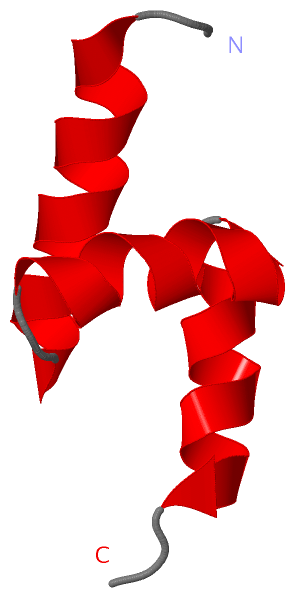
NMR StructureChain A from PDB Type:PROTEIN Length:47 aligned with SWA2_YEAST | Q06677 from UniProtKB/Swiss-Prot Length:668 Alignment length:47 146 156 166 176 SWA2_YEAST 137 ALVDEVKDMEIARLMSLGLSIEEATEFYENDVTYERYLEILKSKQKE 183 SCOP domains d1pgya_ A: Auxilin-like protein Swa2p SCOP domains CATH domains ----------------------------------------------- CATH domains Pfam domains -Ubiq-assoc-1pgyA01 A:2-47 Pfam domains SAPs(SNPs) ----------------------------------------------- SAPs(SNPs) PROSITE ----------------------------------------------- PROSITE Transcript 1 Exon 1.1 PDB: A:1-47 UniProt: 1-668 Transcript 1 1pgy A 1 ALVDEVKDMEIARLMSLGLSIEEATEFYENDVTYERYLEILKSKQKE 47 10 20 30 40
|
||||||||||||||||||||
SCOP Domains (1, 1)
NMR Structure
 |
CATH Domains (0, 0)| (no "CATH Domain" information available for 1PGY) |
Pfam Domains (1, 1)
NMR Structure
|
Gene Ontology (9, 9)|
NMR Structure(hide GO term definitions) Chain A (SWA2_YEAST | Q06677)
|
||||||||||||||||||||||||||||||||||||||||||||||||||||||||||||||||||||||||
Interactive Views
|
||||||||||||||||||||||||||||||||||||||||||||||||||||||||||||||||||||||||||||||||||||||||||||||||||||||||||||||||||||
Still Images
|
||||||||||||||||
Databases
|
||||||||||||||||||||||||||||||||||||||||||||||||||||||||||||||||||||||||||||||||||||||||||||||||||||||||||||||||||||||||||||||||||||||||||||||||||||||||||||||||
Analysis Tools
|
|||||||||||||||||||||||||||||||||||||||||||||||||||||||||||||
Entries Sharing at Least One Protein Chain (UniProt ID)
Related Entries Specified in the PDB File
|
|