Show PDB file:   
         Plain Text   HTML   (compressed file size)
QuickSearch:   
by PDB,NDB,UniProt,PROSITE Code or Search Term(s)  
(-)NMR Structure - model 1
(-)NMR Structure - all models
collapse expand < >
Image NMR Structure - model 1
NMR Structure - model 1  (Jmol Viewer)
Image NMR Structure - all models
NMR Structure - all models  (Jmol Viewer)

(-) Description

Title :  SOLUTION STRUCTURE OF THE DISULPHIDE-LINKED DIMER OF HUMAN INTESTINAL TREFOIL FACTOR (TFF3)
 
Authors :  F. W. Muskett, F. E. May, B. R. Westley, J. Feeney
Date :  21 May 03  (Deposition) - 09 Mar 04  (Release) - 24 Feb 09  (Revision)
Method :  SOLUTION NMR
Resolution :  NOT APPLICABLE
Chains :  NMR Structure  :  1,2  (47x)
Keywords :  Intestinal Trefoil Factor, Itf, Trefoil Factor Family, Tff3, Tff3 Dimer, Trefoil Motif, Solution Structure, Nmr Spectroscopy, Cell Cycle (Keyword Search: [Gene Ontology, PubMed, Web (Google))
 
Reference :  F. W. Muskett, F. E. May, B. R. Westley, J. Feeney
Solution Structure Of The Disulfide-Linked Dimer Of Human Intestinal Trefoil Factor (Tff3): The Intermolecular Orientation And Interactions Are Markedly Different From Those Of Other Dimeric Trefoil Proteins.
Biochemistry V. 42 15139 2003
PubMed-ID: 14690424  |  Reference-DOI: 10.1021/BI030182K
(for further references see the PDB file header)

(-) Compounds

Molecule 1 - TREFOIL FACTOR 3
    Chains1, 2
    EngineeredYES
    Expression SystemESCHERICHIA COLI BL21
    Expression System StrainBL21
    Expression System Taxid511693
    GeneTFF3 OR ITF OR TFI
    Organism CommonHUMAN
    Organism ScientificHOMO SAPIENS
    Organism Taxid9606
    SynonymINTESTINAL TREFOIL FACTOR, HP1.B

 Structural Features

(-) Chains, Units

  
NMR Structure (47x)

Summary Information (see also Sequences/Alignments below)

(-) Ligands, Modified Residues, Ions  (0, 0)

(no "Ligand,Modified Residues,Ions" information available for 1PE3)

(-) Sites  (0, 0)

(no "Site" information available for 1PE3)
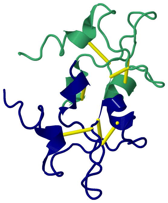
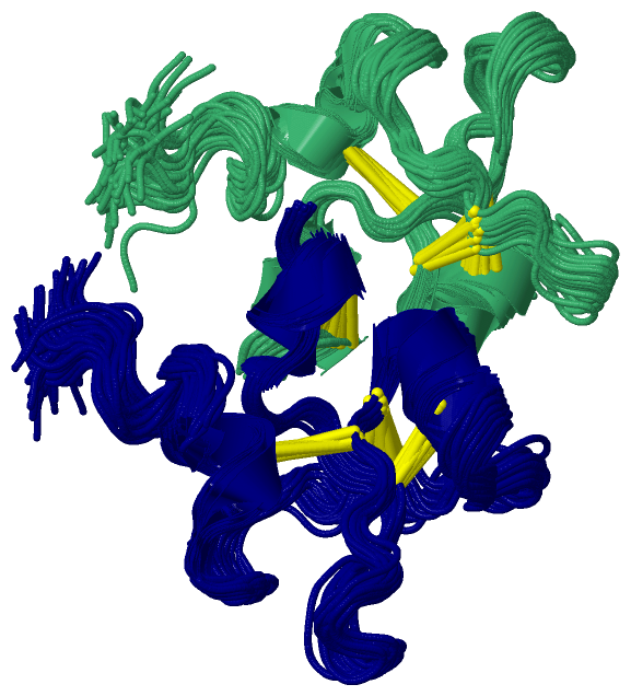
(-) SS Bonds  (7, 7)

NMR Structure
No.Residues
11:11 -1:37
21:21 -1:36
31:31 -1:48
41:57 -2:57
52:11 -2:37
62:21 -2:36
72:31 -2:48

(-) Cis Peptide Bonds  (0, 0)

(no "Cis Peptide Bond" information available for 1PE3)

 Sequence-Structure Mapping

(-) SAPs(SNPs)/Variants  (0, 0)

(no "SAP(SNP)/Variant" information available for 1PE3)

(-) PROSITE Motifs  (2, 4)

NMR Structure (2, 4)
 PROSITEUniProtKBPDB
No.IDACDescriptionIDLocationCountLocation
1P_TREFOIL_2PS51448 P-type 'Trefoil' domain profile.TFF3_HUMAN30-73
 
  21:9-52
2:9-52
2P_TREFOIL_1PS00025 P-type 'Trefoil' domain signature.TFF3_HUMAN39-59
 
  21:18-38
2:18-38

(-) Exons   (0, 0)

(no "Exon" information available for 1PE3)

(-) Sequences/Alignments

NMR Structure
   Reformat: Number of residues per line =  ('0' or empty: single-line sequence representation)
  Number of residues per labelling interval =   
  UniProt sequence: complete  aligned part    
   Show mapping: SCOP domains CATH domains Pfam domains Secondary structure (by author)
SAPs(SNPs) PROSITE motifs Exons
(details for a mapped element are shown in a popup box when the mouse pointer rests over it)
Chain 1 from PDB  Type:PROTEIN  Length:59
 aligned with TFF3_HUMAN | Q07654 from UniProtKB/Swiss-Prot  Length:80

    Alignment length:59
                                    31        41        51        61        71         
            TFF3_HUMAN   22 EEYVGLSANQCAVPAKDRVDCGYPHVTPKECNNRGCCFDSRIPGVPWCFKPLQEAECTF 80
               SCOP domains d1pe31_ 1: Intestinal trefoil factor                        SCOP domains
               CATH domains 1pe3100 1:1-59 Spasmolytic Protein, domain 1                CATH domains
               Pfam domains ----------------------------------------------------------- Pfam domains
         Sec.struct. author ........hhhhh................hhhhhh................hhhhh... Sec.struct. author
                 SAPs(SNPs) ----------------------------------------------------------- SAPs(SNPs)
                PROSITE (1) --------P_TREFOIL_2  PDB: 1:9-52 UniProt: 30-73     ------- PROSITE (1)
                PROSITE (2) -----------------P_TREFOIL_1          --------------------- PROSITE (2)
                 Transcript ----------------------------------------------------------- Transcript
                  1pe3 1  1 EEYVGLSANQCAVPAKDRVDCGYPHVTPKECNNRGCCFDSRIPGVPWCFKPLQEAECTF 59
                                    10        20        30        40        50         

Chain 2 from PDB  Type:PROTEIN  Length:59
 aligned with TFF3_HUMAN | Q07654 from UniProtKB/Swiss-Prot  Length:80

    Alignment length:59
                                    31        41        51        61        71         
            TFF3_HUMAN   22 EEYVGLSANQCAVPAKDRVDCGYPHVTPKECNNRGCCFDSRIPGVPWCFKPLQEAECTF 80
               SCOP domains d1pe32_ 2: Intestinal trefoil factor                        SCOP domains
               CATH domains 1pe3200 2:1-59 Spasmolytic Protein, domain 1                CATH domains
               Pfam domains ----------------------------------------------------------- Pfam domains
         Sec.struct. author ........hhhhh................hhhhhh................hhhhh... Sec.struct. author
                 SAPs(SNPs) ----------------------------------------------------------- SAPs(SNPs)
                PROSITE (1) --------P_TREFOIL_2  PDB: 2:9-52 UniProt: 30-73     ------- PROSITE (1)
                PROSITE (2) -----------------P_TREFOIL_1          --------------------- PROSITE (2)
                 Transcript ----------------------------------------------------------- Transcript
                  1pe3 2  1 EEYVGLSANQCAVPAKDRVDCGYPHVTPKECNNRGCCFDSRIPGVPWCFKPLQEAECTF 59
                                    10        20        30        40        50         

   Legend:   → Mismatch (orange background)
  - → Gap (green background, '-', border residues have a numbering label)
    → Modified Residue (blue background, lower-case, 'x' indicates undefined single-letter code, labelled with number + name)
  x → Chemical Group (purple background, 'x', labelled with number + name, e.g. ACE or NH2)
  extra numbering lines below/above indicate numbering irregularities and modified residue names etc., number ends below/above '|'

 Classification and Annotation

(-) SCOP Domains  (1, 2)

NMR Structure

(-) CATH Domains  (1, 2)

NMR Structure

(-) Pfam Domains  (0, 0)

(no "Pfam Domain" information available for 1PE3)

(-) Gene Ontology  (8, 8)

NMR Structure(hide GO term definitions)
Chain 1,2   (TFF3_HUMAN | Q07654)
molecular function
    GO:0003674    molecular_function    Elemental activities, such as catalysis or binding, describing the actions of a gene product at the molecular level. A given gene product may exhibit one or more molecular functions.
    GO:0005515    protein binding    Interacting selectively and non-covalently with any protein or protein complex (a complex of two or more proteins that may include other nonprotein molecules).
biological process
    GO:0006952    defense response    Reactions, triggered in response to the presence of a foreign body or the occurrence of an injury, which result in restriction of damage to the organism attacked or prevention/recovery from the infection caused by the attack.
    GO:0007586    digestion    The whole of the physical, chemical, and biochemical processes carried out by multicellular organisms to break down ingested nutrients into components that may be easily absorbed and directed into metabolism.
cellular component
    GO:0005737    cytoplasm    All of the contents of a cell excluding the plasma membrane and nucleus, but including other subcellular structures.
    GO:0070062    extracellular exosome    A vesicle that is released into the extracellular region by fusion of the limiting endosomal membrane of a multivesicular body with the plasma membrane. Extracellular exosomes, also simply called exosomes, have a diameter of about 40-100 nm.
    GO:0005576    extracellular region    The space external to the outermost structure of a cell. For cells without external protective or external encapsulating structures this refers to space outside of the plasma membrane. This term covers the host cell environment outside an intracellular parasite.
    GO:0005578    proteinaceous extracellular matrix    A layer consisting mainly of proteins (especially collagen) and glycosaminoglycans (mostly as proteoglycans) that forms a sheet underlying or overlying cells such as endothelial and epithelial cells. The proteins are secreted by cells in the vicinity. An example of this component is found in Mus musculus.

 Visualization

(-) Interactive Views

NMR Structure
  Complete Structure
    Jena3D(integrated viewing of ligand, site, SAP, PROSITE, SCOP information)
    WebMol | AstexViewer[tm]@PDBe
(Java Applets, require no local installation except for Java; loading may be slow)
    STRAP
(Java WebStart application, automatic local installation, requires Java; full application with system access!)
    RasMol
(require local installation)
    Molscript (VRML)
(requires installation of a VRML viewer; select preferred view via VRML and generate a mono or stereo PDF format file)
 
  Ligands, Modified Residues, Ions
(no "Ligands, Modified Residues, Ions" information available for 1pe3)
 
  Sites
(no "Sites" information available for 1pe3)
 
  Cis Peptide Bonds
(no "Cis Peptide Bonds" information available for 1pe3)
 

(-) Still Images

Jmol
  protein: cartoon or spacefill or dots and stick; nucleic acid: cartoon and stick; ligands: spacefill; active site: stick
Molscript
  protein, nucleic acid: cartoon; ligands: spacefill; active site: ball and stick

 Databases and Analysis Tools

(-) Databases

Access by PDB/NDB ID
  1pe3
    Family and Domain InformationProDom | SYSTERS
    General Structural InformationGlycoscienceDB | MMDB | NDB | OCA | PDB | PDBe | PDBj | PDBsum | PDBWiki | PQS | PROTEOPEDIA
    Orientation in MembranesOPM
    Protein SurfaceSURFACE
    Secondary StructureDSSP (structure derived) | HSSP (homology derived)
    Structural GenomicsGeneCensus
    Structural NeighboursCE | VAST
    Structure ClassificationCATH | Dali | SCOP
    Validation and Original DataBMRB Data View | BMRB Restraints Grid | EDS | PROCHECK | RECOORD | WHAT_CHECK
 
Access by UniProt ID/Accession number
  TFF3_HUMAN | Q07654
    Comparative Protein Structure ModelsModBase
    Genomic InformationEnsembl
    Protein-protein InteractionDIP
    Sequence, Family and Domain InformationInterPro | Pfam | SMART | UniProtKB/SwissProt
 
Access by Enzyme Classificator   (EC Number)
  (no 'Enzyme Classificator' available)
    General Enzyme InformationBRENDA | EC-PDB | Enzyme | IntEnz
    PathwayKEGG | MetaCyc
 
Access by Disease Identifier   (MIM ID)
  (no 'MIM ID' available)
    Disease InformationOMIM
 
Access by GenAge ID
  (no 'GenAge ID' available)
    Age Related InformationGenAge

(-) Analysis Tools

Access by PDB/NDB ID
    Domain InformationXDom
    Interatomic Contacts of Structural UnitsCSU
    Ligand-protein ContactsLPC
    Protein CavitiescastP
    Sequence and Secondary StructurePDBCartoon
    Structure AlignmentSTRAP(Java WebStart application, automatic local installation, requires Java; full application with system access!)
    Structure and Sequence BrowserSTING
 
Access by UniProt ID/Accession number
  TFF3_HUMAN | Q07654
    Protein Disorder PredictionDisEMBL | FoldIndex | GLOBPLOT (for more information see DisProt)

 Related Entries

(-) Entries Sharing at Least One Protein Chain (UniProt ID)

UniProtKB/Swiss-Prot
        TFF3_HUMAN | Q076541e9t

(-) Related Entries Specified in the PDB File

1e9t MONOMERIC FORM OF THE PROTEIN