Show PDB file:   
         Plain Text   HTML   (compressed file size)
QuickSearch:   
by PDB,NDB,UniProt,PROSITE Code or Search Term(s)  
(-)NMR Structure - manually
(-)NMR Structure - model 1
(-)NMR Structure - all models
collapse expand < >
Image NMR Structure - manually
NMR Structure - manually  (Jmol Viewer)
Image NMR Structure - model 1
NMR Structure - model 1  (Jmol Viewer)
Image NMR Structure - all models
NMR Structure - all models  (Jmol Viewer)

(-) Description

Title :  THE SOLUTION STRUCTURE OF ANTIFUNGAL PEPTIDE DISTINCT WITH A FIVE-DISULFIDE MOTIF FROM EUCOMMIA ULMOIDES OLIVER
 
Authors :  R. H. Huang, Y. Xiang, G. Z. Tu, Y. Zhang, D. C. Wang
Date :  13 May 03  (Deposition) - 25 May 04  (Release) - 28 Apr 09  (Revision)
Method :  SOLUTION NMR
Resolution :  NOT APPLICABLE
Chains :  NMR Structure  :  A  (21x)
Keywords :  Antifungal Peptide, Chitin-Binding Peptide, Disulfide Stabilized Motif, Antifungal Protein (Keyword Search: [Gene Ontology, PubMed, Web (Google))
 
Reference :  R. H. Huang, Y. Xiang, G. Z. Tu, Y. Zhang, D. C. Wang
Solution Structure Of Eucommia Antifungal Peptide: A Novel Structural Model Distinct With A Five-Disulfide Motif.
Biochemistry V. 43 6005 2004
PubMed-ID: 15147184  |  Reference-DOI: 10.1021/BI036263Y
(for further references see the PDB file header)

(-) Compounds

Molecule 1 - EUCOMMIA ANTIFUNGAL PEPTIDE 2
    ChainsA
    Organism ScientificEUCOMMIA ULMOIDES
    Organism Taxid4392
    Other DetailsEAFP2
    TissueBARK

 Structural Features

(-) Chains, Units

  
NMR Structure (21x)

Summary Information (see also Sequences/Alignments below)

(-) Ligands, Modified Residues, Ions  (1, 1)

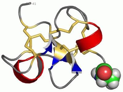
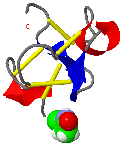
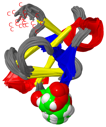
NMR Structure (1, 1)
No.NameCountTypeFull Name
1PCA1Mod. Amino AcidPYROGLUTAMIC ACID

(-) Sites  (0, 0)

(no "Site" information available for 1P9Z)

(-) SS Bonds  (5, 5)

NMR Structure
No.Residues
1A:3 -A:17
2A:7 -A:37
3A:11 -A:23
4A:16 -A:30
5A:35 -A:39

(-) Cis Peptide Bonds  (1, 21)

NMR Structure
No.ModelResidues
11, 2, 3, 4, 5, 6, 7, 8, 9, 10, 11, 12, 13, 14, 15, 16, 17, 18, 19, 20, 21Cys A:7 -Pro A:8

 Sequence-Structure Mapping

(-) SAPs(SNPs)/Variants  (0, 0)

(no "SAP(SNP)/Variant" information available for 1P9Z)

(-) PROSITE Motifs  (2, 2)

NMR Structure (2, 2)
 PROSITEUniProtKBPDB
No.IDACDescriptionIDLocationCountLocation
1CHIT_BIND_I_2PS50941 Chitin-binding type-1 domain profile.EAP2_EUCUL4-41  1A:4-41
2CHIT_BIND_I_1PS00026 Chitin recognition or binding domain signature.EAP2_EUCUL11-30  1A:11-30

(-) Exons   (0, 0)

(no "Exon" information available for 1P9Z)

(-) Sequences/Alignments

NMR Structure
   Reformat: Number of residues per line =  ('0' or empty: single-line sequence representation)
  Number of residues per labelling interval =   
  UniProt sequence: complete  aligned part    
   Show mapping: SCOP domains CATH domains Pfam domains Secondary structure (by author)
SAPs(SNPs) PROSITE motifs Exons
(details for a mapped element are shown in a popup box when the mouse pointer rests over it)
Chain A from PDB  Type:PROTEIN  Length:41
 aligned with EAP2_EUCUL | P83597 from UniProtKB/Swiss-Prot  Length:41

    Alignment length:41
                                    10        20        30        40 
            EAP2_EUCUL    1 QTCASRCPRPCNAGLCCSIYGYCGSGAAYCGAGNCRCQCRG 41
               SCOP domains d1p9za_ A: Antifungal peptide 2           SCOP domains
               CATH domains -1p9zA00 A:2-41                           CATH domains
               Pfam domains -Chitin_bind_1-1p9zA01 A:2-39          -- Pfam domains
         Sec.struct. author ..hhhh.........eee...eee.hhhhh.....ee.... Sec.struct. author
                 SAPs(SNPs) ----------------------------------------- SAPs(SNPs)
                PROSITE (1) ---CHIT_BIND_I_2  PDB: A:4-41             PROSITE (1)
                PROSITE (2) ----------CHIT_BIND_I_1       ----------- PROSITE (2)
                 Transcript ----------------------------------------- Transcript
                  1p9z A  1 xTCASRCPRPCNAGLCCSIYGYCGSGAAYCGAGNCRCQCRG 41
                            |       10        20        30        40 
                            |                                        
                            1-PCA                                    

   Legend:   → Mismatch (orange background)
  - → Gap (green background, '-', border residues have a numbering label)
    → Modified Residue (blue background, lower-case, 'x' indicates undefined single-letter code, labelled with number + name)
  x → Chemical Group (purple background, 'x', labelled with number + name, e.g. ACE or NH2)
  extra numbering lines below/above indicate numbering irregularities and modified residue names etc., number ends below/above '|'

 Classification and Annotation

(-) SCOP Domains  (1, 1)

NMR Structure

(-) CATH Domains  (1, 1)

NMR Structure
(-)
Class: Alpha Beta (26913)

(-) Pfam Domains  (1, 1)

NMR Structure

(-) Gene Ontology  (4, 4)

NMR Structure(hide GO term definitions)
Chain A   (EAP2_EUCUL | P83597)
molecular function
    GO:0008061    chitin binding    Interacting selectively and non-covalently with chitin, a linear polysaccharide consisting of beta-(1->4)-linked N-acetyl-D-glucosamine residues.
biological process
    GO:0006952    defense response    Reactions, triggered in response to the presence of a foreign body or the occurrence of an injury, which result in restriction of damage to the organism attacked or prevention/recovery from the infection caused by the attack.
    GO:0050832    defense response to fungus    Reactions triggered in response to the presence of a fungus that act to protect the cell or organism.
    GO:0031640    killing of cells of other organism    Any process in an organism that results in the killing of cells of another organism, including in some cases the death of the other organism. Killing here refers to the induction of death in one cell by another cell, not cell-autonomous death due to internal or other environmental conditions.

 Visualization

(-) Interactive Views

NMR Structure
  Complete Structure
    Jena3D(integrated viewing of ligand, site, SAP, PROSITE, SCOP information)
    WebMol | AstexViewer[tm]@PDBe
(Java Applets, require no local installation except for Java; loading may be slow)
    STRAP
(Java WebStart application, automatic local installation, requires Java; full application with system access!)
    RasMol
(require local installation)
    Molscript (VRML)
(requires installation of a VRML viewer; select preferred view via VRML and generate a mono or stereo PDF format file)
 
  Ligands, Modified Residues, Ions
    PCA  [ RasMol | Jena3D ]  +environment [ RasMol | Jena3D ]
 
  Sites
(no "Sites" information available for 1p9z)
 
  Cis Peptide Bonds
    Cys A:7 - Pro A:8   [ RasMol ]  
 

(-) Still Images

Jmol
  protein: cartoon or spacefill or dots and stick; nucleic acid: cartoon and stick; ligands: spacefill; active site: stick
Molscript
  protein, nucleic acid: cartoon; ligands: spacefill; active site: ball and stick

 Databases and Analysis Tools

(-) Databases

Access by PDB/NDB ID
  1p9z
    Family and Domain InformationProDom | SYSTERS
    General Structural InformationGlycoscienceDB | MMDB | NDB | OCA | PDB | PDBe | PDBj | PDBsum | PDBWiki | PQS | PROTEOPEDIA
    Orientation in MembranesOPM
    Protein SurfaceSURFACE
    Secondary StructureDSSP (structure derived) | HSSP (homology derived)
    Structural GenomicsGeneCensus
    Structural NeighboursCE | VAST
    Structure ClassificationCATH | Dali | SCOP
    Validation and Original DataBMRB Data View | BMRB Restraints Grid | EDS | PROCHECK | RECOORD | WHAT_CHECK
 
Access by UniProt ID/Accession number
  EAP2_EUCUL | P83597
    Comparative Protein Structure ModelsModBase
    Genomic InformationEnsembl
    Protein-protein InteractionDIP
    Sequence, Family and Domain InformationInterPro | Pfam | SMART | UniProtKB/SwissProt
 
Access by Enzyme Classificator   (EC Number)
  (no 'Enzyme Classificator' available)
    General Enzyme InformationBRENDA | EC-PDB | Enzyme | IntEnz
    PathwayKEGG | MetaCyc
 
Access by Disease Identifier   (MIM ID)
  (no 'MIM ID' available)
    Disease InformationOMIM
 
Access by GenAge ID
  (no 'GenAge ID' available)
    Age Related InformationGenAge

(-) Analysis Tools

Access by PDB/NDB ID
    Domain InformationXDom
    Interatomic Contacts of Structural UnitsCSU
    Ligand-protein ContactsLPC
    Protein CavitiescastP
    Sequence and Secondary StructurePDBCartoon
    Structure AlignmentSTRAP(Java WebStart application, automatic local installation, requires Java; full application with system access!)
    Structure and Sequence BrowserSTING
 
Access by UniProt ID/Accession number
  EAP2_EUCUL | P83597
    Protein Disorder PredictionDisEMBL | FoldIndex | GLOBPLOT (for more information see DisProt)

 Related Entries

(-) Entries Sharing at Least One Protein Chain (UniProt ID)

UniProtKB/Swiss-Prot
        EAP2_EUCUL | P835971p9g

(-) Related Entries Specified in the PDB File

5795 THE CHEMICAL SHIFTS OF THE HYDROGENS