Show PDB file:   
         Plain Text   HTML   (compressed file size)
QuickSearch:   
by PDB,NDB,UniProt,PROSITE Code or Search Term(s)  
(-)Asym./Biol. Unit
(-)Asym./Biol. Unit - sites
collapse expand < >
Image Asym./Biol. Unit
Asym./Biol. Unit  (Jmol Viewer)
Image Asym./Biol. Unit - sites
Asym./Biol. Unit - sites  (Jmol Viewer)

(-) Description

Title :  1.7A CRYSTAL STRUCTURE OF PROTEIN PG110 FROM STAPHYLOCOCCUS AUREUS
 
Authors :  R. Zhang, M. Zhou, G. Joachimiak, O. Schneewind, A. Joachimiak, Midwest Center For Structural Genomics (Mcsg)
Date :  09 May 03  (Deposition) - 20 Jan 04  (Release) - 24 Feb 09  (Revision)
Method :  X-RAY DIFFRACTION
Resolution :  1.70
Chains :  Asym./Biol. Unit :  A
Keywords :  Structural Genomics, Psi, Protein Structure Initiative, Midwest Center For Structural Genomics, Mcsg, Unknown Function (Keyword Search: [Gene Ontology, PubMed, Web (Google))
 
Reference :  W. A. Williams, R. G. Zhang, M. Zhou, G. Joachimiak, P. Gornicki, D. Missiakas, A. Joachimiak
The Membrane-Associated Lipoprotein-9 Gmpc From Staphylococcus Aureus Binds The Dipeptide Glymet Via Side Chain Interactions.
Biochemistry V. 43 16193 2004
PubMed-ID: 15610013  |  Reference-DOI: 10.1021/BI048877O
(for further references see the PDB file header)

(-) Compounds

Molecule 1 - HYPOTHETICAL PROTEIN PG110
    ChainsA
    EngineeredYES
    Expression SystemESCHERICHIA COLI BL21(DE3)
    Expression System PlasmidPDM 68
    Expression System StrainBL21 (DE3)
    Expression System Taxid469008
    Expression System Vector TypePLASMID
    FragmentRESIDUES 19-280
    GeneSA0422
    Organism ScientificSTAPHYLOCOCCUS AUREUS SUBSP. AUREUS MU50
    Organism Taxid158878
    StrainSUBSP. AUREUS MU50

 Structural Features

(-) Chains, Units

  1
Asymmetric/Biological Unit A

Summary Information (see also Sequences/Alignments below)
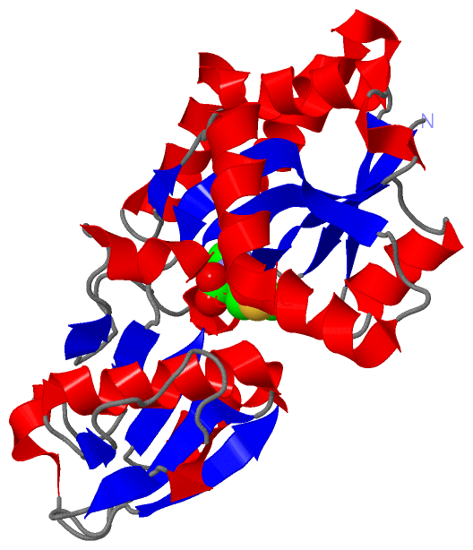
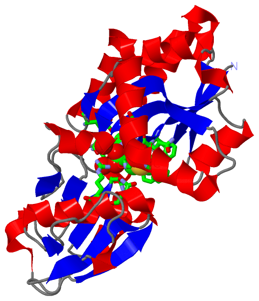
(-) Ligands, Modified Residues, Ions  (2, 2)

Asymmetric/Biological Unit (2, 2)
No.NameCountTypeFull Name
1GLY1Mod. Amino AcidGLYCINE
2MET1Mod. Amino AcidMETHIONINE

(-) Sites  (2, 2)

Asymmetric Unit (2, 2)
No.NameEvidenceResiduesDescription
1AC1SOFTWARESER A:47 , ALA A:119 , ASN A:208 , GLY A:209 , ASN A:238 , GLU A:276 , MET A:301BINDING SITE FOR RESIDUE GLY A 300
2AC2SOFTWARETYR A:75 , PHE A:92 , GLN A:93 , HIS A:94 , PHE A:97 , ASN A:148 , ARG A:151 , ASN A:207 , ASN A:238 , GLY A:300 , HOH A:404 , HOH A:405BINDING SITE FOR RESIDUE MET A 301

(-) SS Bonds  (0, 0)

(no "SS Bond" information available for 1P99)

(-) Cis Peptide Bonds  (0, 0)

(no "Cis Peptide Bond" information available for 1P99)

 Sequence-Structure Mapping

(-) SAPs(SNPs)/Variants  (0, 0)

(no "SAP(SNP)/Variant" information available for 1P99)

(-) PROSITE Motifs  (0, 0)

(no "PROSITE Motif" information available for 1P99)

(-) Exons   (0, 0)

(no "Exon" information available for 1P99)

(-) Sequences/Alignments

Asymmetric/Biological Unit
   Reformat: Number of residues per line =  ('0' or empty: single-line sequence representation)
  Number of residues per labelling interval =   
  UniProt sequence: complete  aligned part    
   Show mapping: SCOP domains CATH domains Pfam domains Secondary structure (by author)
SAPs(SNPs) PROSITE motifs Exons
(details for a mapped element are shown in a popup box when the mouse pointer rests over it)
Chain A from PDB  Type:PROTEIN  Length:257
 aligned with A0A0H3JTG5_S | A0A0H3JTG5 from UniProtKB/TrEMBL  Length:280

    Alignment length:258
                                                                                                                                                                                                                                                                                         280  
                                    34        44        54        64        74        84        94       104       114       124       134       144       154       164       174       184       194       204       214       224       234       244       254       264       274     |  
         A0A0H3JTG5_S    25 KVTIGVASNDTKAWEKVKELAKKDDIDVEIKHFSDYNLPNKALNDGDIDMNAFQHFAFLDQYKKAHKGTKISALSTTVLAPLGIYSDKIKDVKKVKDGAKVVIPNDVSNQARALKLLEAAGLIKLKKDFGLAGTVKDITSNPKHLKITAVDAQQTARALSDVDIAVINNGVATKAGKDPKNDPIFLEKSNSDAVKPYINIVAVNDKDLDNKTYAKIVELYHSKEAQKALQEDVKDGEKPVNLSKDEIKAIETSLAK--   -
               SCOP domains d1p99a_ A: Putative lipoprotein (NlpA family)                                                                                                                                                                                                                      SCOP domains
               CATH domains 1p99A01 A:40-118,A:230-294 Periplasmic binding protein-like II                 1p99A02 A:119-229 Periplasmic binding protein-like II                                                          1p99A01 A:40-118,A:230-294 Periplasmic binding protein-like II   --- CATH domains
               Pfam domains ------------------------------------------------------------------------------------------------------------------------------------------------------------------------------------------------------------------------------------------------------------------ Pfam domains
         Sec.struct. author .eeeee....hhhhhhhhhhhhhhh..eeeee.....hhhhhhhh....eeeeeehhhhhhhhhhh....eeeeeeeee...eee.....hhhhh....eeeee.hhhhhhhhhhhhhhh...ee........hhh.eee.....eeeeehhhhhhhhhhh..eeeehhhhhhhh.........ee....hhhhhh.eeeeeee.hhh.hhhhhhhhhhhhhhhhhhhhhhhhh...ee...hhhhhhhhhhhhh-.. Sec.struct. author
                 SAPs(SNPs) ------------------------------------------------------------------------------------------------------------------------------------------------------------------------------------------------------------------------------------------------------------------ SAPs(SNPs)
                    PROSITE ------------------------------------------------------------------------------------------------------------------------------------------------------------------------------------------------------------------------------------------------------------------ PROSITE
                 Transcript ------------------------------------------------------------------------------------------------------------------------------------------------------------------------------------------------------------------------------------------------------------------ Transcript
                 1p99 A  40 KVTIGVASNDTKAWEKVKELAKKDDIDVEIKHFSDYNLPNKALNDGDIDMNAFQHFAFLDQYKKAHKGTKISALSTTVLAPLGIYSDKIKDVKKVKDGAKVVIPNDVSNQARALKLLEAAGLIKLKKDFGLAGTVKDITSNPKHLKITAVDAQQTARALSDVDIAVINNGVATKAGKDPKNDPIFLEKSNSDAVKPYINIVAVNDKDLDNKTYAKIVELYHSKEAQKALQEDVKDGEKPVNLSKDEIKAIETSLA-GM 301
                                    49        59        69        79        89        99       109       119       129       139       149       159       169       179       189       199       209       219       229       239       249       259       269       279       289    | | 
                                                                                                                                                                                                                                                                                        294 | 
                                                                                                                                                                                                                                                                                          300 

   Legend:   → Mismatch (orange background)
  - → Gap (green background, '-', border residues have a numbering label)
    → Modified Residue (blue background, lower-case, 'x' indicates undefined single-letter code, labelled with number + name)
  x → Chemical Group (purple background, 'x', labelled with number + name, e.g. ACE or NH2)
  extra numbering lines below/above indicate numbering irregularities and modified residue names etc., number ends below/above '|'

 Classification and Annotation

(-) SCOP Domains  (1, 1)

Asymmetric/Biological Unit

(-) CATH Domains  (1, 2)

Asymmetric/Biological Unit
(-)
Class: Alpha Beta (26913)

(-) Pfam Domains  (0, 0)

(no "Pfam Domain" information available for 1P99)

(-) Gene Ontology  (0, 0)

Asymmetric/Biological Unit(hide GO term definitions)
    (no "Gene Ontology" information available for 1P99)

 Visualization

(-) Interactive Views

Asymmetric/Biological Unit
  Complete Structure
    Jena3D(integrated viewing of ligand, site, SAP, PROSITE, SCOP information)
    WebMol | AstexViewer[tm]@PDBe
(Java Applets, require no local installation except for Java; loading may be slow)
    STRAP
(Java WebStart application, automatic local installation, requires Java; full application with system access!)
    RasMol
(require local installation)
    Molscript (VRML)
(requires installation of a VRML viewer; select preferred view via VRML and generate a mono or stereo PDF format file)
 
  Ligands, Modified Residues, Ions
    GLY  [ RasMol | Jena3D ]  +environment [ RasMol | Jena3D ]
    MET  [ RasMol | Jena3D ]  +environment [ RasMol | Jena3D ]
 
  Sites
    AC1  [ RasMol ]  +environment [ RasMol ]
    AC2  [ RasMol ]  +environment [ RasMol ]
 
  Cis Peptide Bonds
(no "Cis Peptide Bonds" information available for 1p99)
 

(-) Still Images

Jmol
  protein: cartoon or spacefill or dots and stick; nucleic acid: cartoon and stick; ligands: spacefill; active site: stick
Molscript
  protein, nucleic acid: cartoon; ligands: spacefill; active site: ball and stick

 Databases and Analysis Tools

(-) Databases

Access by PDB/NDB ID
  1p99
    Family and Domain InformationProDom | SYSTERS
    General Structural InformationGlycoscienceDB | MMDB | NDB | OCA | PDB | PDBe | PDBj | PDBsum | PDBWiki | PQS | PROTEOPEDIA
    Orientation in MembranesOPM
    Protein SurfaceSURFACE
    Secondary StructureDSSP (structure derived) | HSSP (homology derived)
    Structural GenomicsGeneCensus
    Structural NeighboursCE | VAST
    Structure ClassificationCATH | Dali | SCOP
    Validation and Original DataBMRB Data View | BMRB Restraints Grid | EDS | PROCHECK | RECOORD | WHAT_CHECK
 
Access by UniProt ID/Accession number
  A0A0H3JTG5_S | A0A0H3JTG5
    Comparative Protein Structure ModelsModBase
    Genomic InformationEnsembl
    Protein-protein InteractionDIP
    Sequence, Family and Domain InformationInterPro | Pfam | SMART | UniProtKB/TrEMBL
 
Access by Enzyme Classificator   (EC Number)
  (no 'Enzyme Classificator' available)
    General Enzyme InformationBRENDA | EC-PDB | Enzyme | IntEnz
    PathwayKEGG | MetaCyc
 
Access by Disease Identifier   (MIM ID)
  (no 'MIM ID' available)
    Disease InformationOMIM
 
Access by GenAge ID
  (no 'GenAge ID' available)
    Age Related InformationGenAge

(-) Analysis Tools

Access by PDB/NDB ID
    Domain InformationXDom
    Interatomic Contacts of Structural UnitsCSU
    Ligand-protein ContactsLPC
    Protein CavitiescastP
    Sequence and Secondary StructurePDBCartoon
    Structure AlignmentSTRAP(Java WebStart application, automatic local installation, requires Java; full application with system access!)
    Structure and Sequence BrowserSTING
 
Access by UniProt ID/Accession number
  A0A0H3JTG5_S | A0A0H3JTG5
    Protein Disorder PredictionDisEMBL | FoldIndex | GLOBPLOT (for more information see DisProt)

 Related Entries

(-) Entries Sharing at Least One Protein Chain (UniProt ID)

(no "Entries Sharing at Least One Protein Chain" available for 1P99)

(-) Related Entries Specified in the PDB File

(no "Related Entries Specified in the PDB File" available for 1P99)