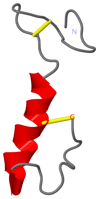
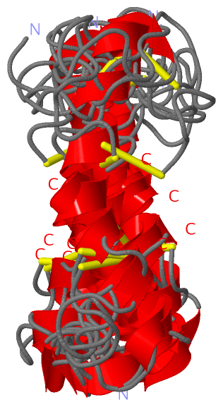
Show PDB file:   
         Plain Text   HTML   (compressed file size)
QuickSearch:   
by PDB,NDB,UniProt,PROSITE Code or Search Term(s)  
(-)NMR Structure - model 1
(-)NMR Structure - all models
collapse expand < >
Image NMR Structure - model 1
NMR Structure - model 1  (Jmol Viewer)
Image NMR Structure - all models
NMR Structure - all models  (Jmol Viewer)

(-) Description

Title :  SAKACIN P VARIANT THAT IS STRUCTURALLY STABILIZED BY AN INSERTED C-TERMINAL DISULFIDE BRIDGE.
 
Authors :  M. Uteng, H. H. Hauge, P. R. Markwick, G. Fimland, D. Mantzilas, J. Nissen-Meyer, C. Muhle-Goll
Date :  28 May 03  (Deposition) - 22 Sep 03  (Release) - 24 Feb 09  (Revision)
Method :  SOLUTION NMR
Resolution :  NOT APPLICABLE
Chains :  NMR Structure  :  A  (10x)
Keywords :  Antibiotic, Pediocin-Like Bacteriocins, Antimicrobial Peptides, Sakacin Antibiotic, Bacteriocin (Keyword Search: [Gene Ontology, PubMed, Web (Google))
 
Reference :  M. Uteng, H. H. Hauge, P. R. Markwick, G. Fimland, D. Mantzilas, J. Nissen-Meyer, C. Muhle-Goll
Three-Dimensional Structure In Lipid Micelles Of The Pediocin-Like Antimicrobial Peptide Sakacin P And A Sakacin P Variant That Is Structurally Stabilized By An Inserted C-Terminal Disulfide Bridge
Biochemistry V. 42 11417 2003
PubMed-ID: 14516192  |  Reference-DOI: 10.1021/BI034572I

(-) Compounds

Molecule 1 - BACTERIOCIN SAKACIN P
    ChainsA
    EngineeredYES
    Expression SystemLACTOBACILLUS SAKE
    Expression System PlasmidPGF10
    Expression System StrainLB790
    Expression System Taxid1599
    Expression System VariantLB790/PGF10
    Expression System VectorPMG36E
    MutationYES
    Organism ScientificLACTOBACILLUS SAKE
    Organism Taxid1599
    Other DetailsSAKACIN P VARIANT THAT IS STRUCTURALLY STABILIZED BY AN INSERTED C-TERMINAL DISULFIDE BRIDGE.
    StrainLB790
    SynonymSAKACIN 674
    VariantLB790/PGF10

 Structural Features

(-) Chains, Units

  
NMR Structure (10x)

Summary Information (see also Sequences/Alignments below)

(-) Ligands, Modified Residues, Ions  (0, 0)

(no "Ligand,Modified Residues,Ions" information available for 1OHM)

(-) Sites  (0, 0)

(no "Site" information available for 1OHM)

(-) SS Bonds  (2, 2)

NMR Structure
No.Residues
1A:9 -A:14
2A:24 -A:44

(-) Cis Peptide Bonds  (4, 4)

NMR Structure
No.ModelResidues
12Cys A:9 -Gly A:10
25Thr A:15 -Val A:16
35Asn A:32 -Trp A:33
410His A:12 -Ser A:13

 Sequence-Structure Mapping

(-) SAPs(SNPs)/Variants  (0, 0)

(no "SAP(SNP)/Variant" information available for 1OHM)

(-) PROSITE Motifs  (1, 1)

NMR Structure (1, 1)
 PROSITEUniProtKBPDB
No.IDACDescriptionIDLocationCountLocation
1BACTERIOCIN_IIAPS60030 Bacteriocin class IIa family signature.SAKP_LACSK21-32  1A:3-14

(-) Exons   (0, 0)

(no "Exon" information available for 1OHM)

(-) Sequences/Alignments

NMR Structure
   Reformat: Number of residues per line =  ('0' or empty: single-line sequence representation)
  Number of residues per labelling interval =   
  UniProt sequence: complete  aligned part    
   Show mapping: SCOP domains CATH domains Pfam domains Secondary structure (by author)
SAPs(SNPs) PROSITE motifs Exons
(details for a mapped element are shown in a popup box when the mouse pointer rests over it)
Chain A from PDB  Type:PROTEIN  Length:44
 aligned with SAKP_LACSK | P35618 from UniProtKB/Swiss-Prot  Length:61

    Alignment length:44
                                                                     61 
                                    28        38        48        58  | 
            SAKP_LACSK   19 KYYGNGVHCGKHSCTVDWGTAIGNIGNNAAANWATGGNAGWNK-  -
               SCOP domains d1ohma_ A: Sakacin P                         SCOP domains
               CATH domains -------------------------------------------- CATH domains
               Pfam domains Bacteriocin_II-1ohmA01 A:1-36       -------- Pfam domains
         Sec.struct. author .................hhhhhhhhhhhhhhhh........... Sec.struct. author
                 SAPs(SNPs) -------------------------------------------- SAPs(SNPs)
                    PROSITE --BACTERIOCIN_------------------------------ PROSITE
                 Transcript -------------------------------------------- Transcript
                  1ohm A  1 KYYGNGVHCGKHSCTVDWGTAIGCIGNNAAANWATGGNAGWNKC 44
                                    10        20        30        40    

   Legend:   → Mismatch (orange background)
  - → Gap (green background, '-', border residues have a numbering label)
    → Modified Residue (blue background, lower-case, 'x' indicates undefined single-letter code, labelled with number + name)
  x → Chemical Group (purple background, 'x', labelled with number + name, e.g. ACE or NH2)
  extra numbering lines below/above indicate numbering irregularities and modified residue names etc., number ends below/above '|'

 Classification and Annotation

(-) SCOP Domains  (1, 1)

NMR Structure
(-)
Class: Peptides (792)

(-) CATH Domains  (0, 0)

(no "CATH Domain" information available for 1OHM)

(-) Pfam Domains  (1, 1)

NMR Structure

(-) Gene Ontology  (3, 3)

NMR Structure(hide GO term definitions)
Chain A   (SAKP_LACSK | P35618)
biological process
    GO:0019835    cytolysis    The rupture of cell membranes and the loss of cytoplasm.
    GO:0042742    defense response to bacterium    Reactions triggered in response to the presence of a bacterium that act to protect the cell or organism.
cellular component
    GO:0005576    extracellular region    The space external to the outermost structure of a cell. For cells without external protective or external encapsulating structures this refers to space outside of the plasma membrane. This term covers the host cell environment outside an intracellular parasite.

 Visualization

(-) Interactive Views

NMR Structure
  Complete Structure
    Jena3D(integrated viewing of ligand, site, SAP, PROSITE, SCOP information)
    WebMol | AstexViewer[tm]@PDBe
(Java Applets, require no local installation except for Java; loading may be slow)
    STRAP
(Java WebStart application, automatic local installation, requires Java; full application with system access!)
    RasMol
(require local installation)
    Molscript (VRML)
(requires installation of a VRML viewer; select preferred view via VRML and generate a mono or stereo PDF format file)
 
  Ligands, Modified Residues, Ions
(no "Ligands, Modified Residues, Ions" information available for 1ohm)
 
  Sites
(no "Sites" information available for 1ohm)
 
  Cis Peptide Bonds
    Asn A:32 - Trp A:33   [ RasMol ]  
    Cys A:9 - Gly A:10   [ RasMol ]  
    His A:12 - Ser A:13   [ RasMol ]  
    Thr A:15 - Val A:16   [ RasMol ]  
 

(-) Still Images

Jmol
  protein: cartoon or spacefill or dots and stick; nucleic acid: cartoon and stick; ligands: spacefill; active site: stick
Molscript
  protein, nucleic acid: cartoon; ligands: spacefill; active site: ball and stick

 Databases and Analysis Tools

(-) Databases

Access by PDB/NDB ID
  1ohm
    Family and Domain InformationProDom | SYSTERS
    General Structural InformationGlycoscienceDB | MMDB | NDB | OCA | PDB | PDBe | PDBj | PDBsum | PDBWiki | PQS | PROTEOPEDIA
    Orientation in MembranesOPM
    Protein SurfaceSURFACE
    Secondary StructureDSSP (structure derived) | HSSP (homology derived)
    Structural GenomicsGeneCensus
    Structural NeighboursCE | VAST
    Structure ClassificationCATH | Dali | SCOP
    Validation and Original DataBMRB Data View | BMRB Restraints Grid | EDS | PROCHECK | RECOORD | WHAT_CHECK
 
Access by UniProt ID/Accession number
  SAKP_LACSK | P35618
    Comparative Protein Structure ModelsModBase
    Genomic InformationEnsembl
    Protein-protein InteractionDIP
    Sequence, Family and Domain InformationInterPro | Pfam | SMART | UniProtKB/SwissProt
 
Access by Enzyme Classificator   (EC Number)
  (no 'Enzyme Classificator' available)
    General Enzyme InformationBRENDA | EC-PDB | Enzyme | IntEnz
    PathwayKEGG | MetaCyc
 
Access by Disease Identifier   (MIM ID)
  (no 'MIM ID' available)
    Disease InformationOMIM
 
Access by GenAge ID
  (no 'GenAge ID' available)
    Age Related InformationGenAge

(-) Analysis Tools

Access by PDB/NDB ID
    Domain InformationXDom
    Interatomic Contacts of Structural UnitsCSU
    Ligand-protein ContactsLPC
    Protein CavitiescastP
    Sequence and Secondary StructurePDBCartoon
    Structure AlignmentSTRAP(Java WebStart application, automatic local installation, requires Java; full application with system access!)
    Structure and Sequence BrowserSTING
 
Access by UniProt ID/Accession number
  SAKP_LACSK | P35618
    Protein Disorder PredictionDisEMBL | FoldIndex | GLOBPLOT (for more information see DisProt)

 Related Entries

(-) Entries Sharing at Least One Protein Chain (UniProt ID)

UniProtKB/Swiss-Prot
        SAKP_LACSK | P356181og7 1ohn

(-) Related Entries Specified in the PDB File

1og7 THREE-DIMENSIONAL STRUCTURE IN LIPID MICELLES OF THE PEDIOCIN-LIKE ANTIMICROBIAL PEPTIDE SAKACIN P.
1ohn THREE-DIMENSIONAL STRUCTURE IN LIPID MICELLES OF THE PEDIOCIN-LIKE ANTIMICROBIAL PEPTIDE SAKACIN P.