|
|
|
|
 Description
|
|
Compounds
|
||||||||||||||||||||||||||||||||||||||||
Chains, Units
Summary Information (see also Sequences/Alignments below) |
Ligands, Modified Residues, Ions (11, 12)
Asymmetric/Biological Unit (11, 12)
|
Sites (2, 2)
Asymmetric Unit (2, 2)
|
SS Bonds (0, 0)| (no "SS Bond" information available for 1OFM) |
Cis Peptide Bonds (1, 1)
Asymmetric/Biological Unit
|
||||||||
SAPs(SNPs)/Variants (0, 0)| (no "SAP(SNP)/Variant" information available for 1OFM) |
PROSITE Motifs (0, 0)| (no "PROSITE Motif" information available for 1OFM) |
Exons (0, 0)| (no "Exon" information available for 1OFM) |
Sequences/Alignments
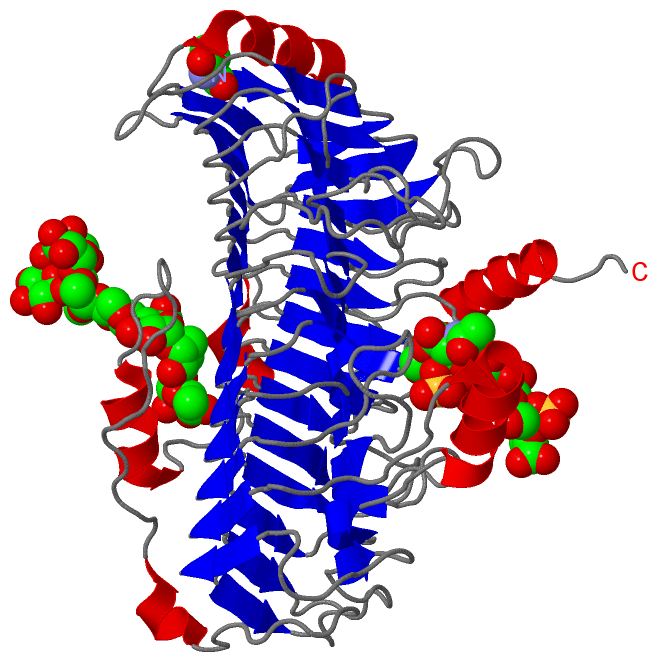
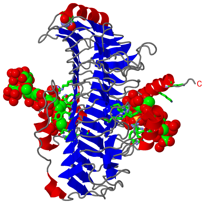
Asymmetric/Biological UnitChain A from PDB Type:PROTEIN Length:481 aligned with CSLB_PEDHD | Q46079 from UniProtKB/Swiss-Prot Length:506 Alignment length:481 35 45 55 65 75 85 95 105 115 125 135 145 155 165 175 185 195 205 215 225 235 245 255 265 275 285 295 305 315 325 335 345 355 365 375 385 395 405 415 425 435 445 455 465 475 485 495 505 CSLB_PEDHD 26 QVVASNETLYQVVKEVKPGGLVQIADGTYKDVQLIVSNSGKSGLPITIKALNPGKVFFTGDAKVELRGEHLILEGIWFKDGNRAIQAWKSHGPGLVAIYGSYNRITACVFDCFDEANSAYITTSLTEDGKVPQHCRIDHCSFTDKITFDQVINLNNTARAIKDGSVGGPAMYHRVDHCFFSNPQKPGNAGGGIRIGYYRNDIGRCLVDSNLFMRQDSEAEIITSKSQENVYYGNTYLNCQGTMNFRHGDHQVAINNFYIGNDQRFGYGGMFVWGSRHVIACNYFELSETIKSRGNAALYLNPGAMASEHALAFDMLIANNAFINVNGYAIHFNPLDERRKEYCAANRLKFETPHQLMLKGNLFFKDKPYVYPFFKDDYFIAGKNSWTGNVALGVEKGIPVNISANRSAYKPVKIKDIQPIEGIALDLNALISKGITGKPLSWDEVRPYWLKEMPGTYALTARLSADRAAKFKAVIKRNKEH 506 SCOP domains d1ofma_ A: Chondroitinase B SCOP domains CATH domains -1ofmA00 A:27-506 Single-stranded right-handed beta-helix, Pectin lyase-like CATH domains Pfam domains ------------------------------------------------------------------------------------------------------------------------------------------------------------------------------------------------------------------------------------------------------------------------------------------------------------------------------------------------------------------------------------------------------------------------------------------------------------------------------------------------- Pfam domains SAPs(SNPs) ------------------------------------------------------------------------------------------------------------------------------------------------------------------------------------------------------------------------------------------------------------------------------------------------------------------------------------------------------------------------------------------------------------------------------------------------------------------------------------------------- SAPs(SNPs) PROSITE ------------------------------------------------------------------------------------------------------------------------------------------------------------------------------------------------------------------------------------------------------------------------------------------------------------------------------------------------------------------------------------------------------------------------------------------------------------------------------------------------- PROSITE Transcript ------------------------------------------------------------------------------------------------------------------------------------------------------------------------------------------------------------------------------------------------------------------------------------------------------------------------------------------------------------------------------------------------------------------------------------------------------------------------------------------------- Transcript 1ofm A 26 xVVASNETLYQVVKEVKPGGLVQIADGTYKDVQLIVSNSGKSGLPITIKALNPGKVFFTGDAKVELRGEHLILEGIWFKDGNRAIQAWKSHGPGLVAIYGSYNRITACVFDCFDEANSAYITTSLTEDGKVPQHCRIDHCSFTDKITFDQVINLNNTARAIKDGSVGGPAMYHRVDHCFFSNPQKPGNAGGGIRIGYYRNDIGRCLVDSNLFMRQDSEAEIITSKSQENVYYGNTYLNCQGTMNFRHGDHQVAINNFYIGNDQRFGYGGMFVWGSRHVIACNYFELSETIKSRGNAALYLNPGAMASEHALAFDMLIANNAFINVNGYAIHFNPLDERRKEYCAANRLKFETPHQLMLKGNLFFKDKPYVYPFFKDDYFIAGKNSWTGNVALGVEKGIPVNISANRSAYKPVKIKDIQPIEGIALDLNALISKGITGKPLSWDEVRPYWLKEMPGTYALTARLSADRAAKFKAVIKRNKEH 506 | 35 45 55 65 75 85 95 105 115 125 135 145 155 165 175 185 195 205 215 225 235 245 255 265 275 285 295 305 315 325 335 345 355 365 375 385 395 405 415 425 435 445 455 465 475 485 495 505 | 26-PCA
|
||||||||||||||||||||
SCOP Domains (1, 1)
Asymmetric/Biological Unit
 |
CATH Domains (1, 1)
Asymmetric/Biological Unit
|
Pfam Domains (0, 0)| (no "Pfam Domain" information available for 1OFM) |
Gene Ontology (2, 2)|
Asymmetric/Biological Unit(hide GO term definitions) Chain A (CSLB_PEDHD | Q46079)
|
||||||||||||||||||
Interactive Views
|
||||||||||||||||||||||||||||||||||||||||||||||||||||||||||||||||||||||||||||||||||||||||||||||||||||||||||||||||||||||||||||||||||||||||||||||||||||||||||||||||||||||||||||||||||||||||||||||||||||
Still Images
|
||||||||||||||||
Databases
|
||||||||||||||||||||||||||||||||||||||||||||||||||||||||||||||||||||||||||||||||||||||||||||||||||||||||||||||||||||||||||||||||||||||||||||||||||||||||||||||||
Analysis Tools
|
|||||||||||||||||||||||||||||||||||||||||||||||||||||||||||||
Entries Sharing at Least One Protein Chain (UniProt ID)
Related Entries Specified in the PDB File
|
|