Show PDB file:   
         Plain Text   HTML   (compressed file size)
QuickSearch:   
by PDB,NDB,UniProt,PROSITE Code or Search Term(s)  
(-)Asym./Biol. Unit
(-)Asym./Biol. Unit - sites
collapse expand < >
Image Asym./Biol. Unit
Asym./Biol. Unit  (Jmol Viewer)
Image Asym./Biol. Unit - sites
Asym./Biol. Unit - sites  (Jmol Viewer)

(-) Description

Title :  STRUCTURE OF MELANOCARPUS ALBOMYCES ENDOGLUCANASE IN COMPLEX WITH CELLOBIOSE
 
Authors :  M. Hirvonen, A. C. Papageorgiou
Date :  02 Jan 03  (Deposition) - 29 May 03  (Release) - 24 Feb 09  (Revision)
Method :  X-RAY DIFFRACTION
Resolution :  2.00
Chains :  Asym./Biol. Unit :  A
Keywords :  Hydrolase, Cellulase, Cellulose Degradation, Glycoside Hydrolases (Keyword Search: [Gene Ontology, PubMed, Web (Google))
 
Reference :  M. Hirvonen, A. C. Papageorgiou
Crystal Structure Of A Family 45 Endoglucanase From Melanocarpus Albomyces: Mechanistic Implications Based On The Free And Cellobiose-Bound Forms
J. Mol. Biol. V. 329 403 2003
PubMed-ID: 12767825  |  Reference-DOI: 10.1016/S0022-2836(03)00467-4

(-) Compounds

Molecule 1 - CELLULASE
    ChainsA
    EC Number3.2.1.4
    Organism ScientificMELANOCARPUS ALBOMYCES
    Organism Taxid204285
    SynonymENDOGLUCANASE

 Structural Features

(-) Chains, Units

  1
Asymmetric/Biological Unit A

Summary Information (see also Sequences/Alignments below)

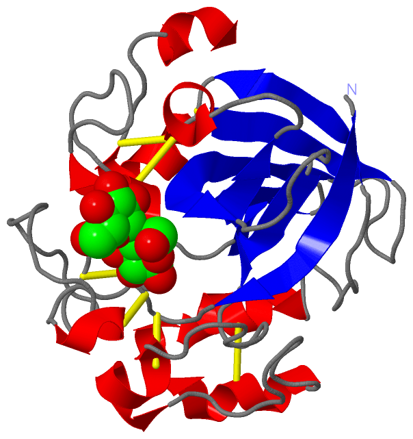
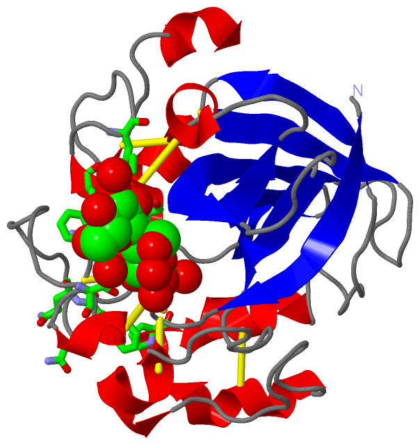
(-) Ligands, Modified Residues, Ions  (1, 1)

Asymmetric/Biological Unit (1, 1)
No.NameCountTypeFull Name
1CBI1Ligand/IonCELLOBIOSE

(-) Sites  (1, 1)

Asymmetric Unit (1, 1)
No.NameEvidenceResiduesDescription
1AC1SOFTWARECYS A:12 , LYS A:13 , SER A:15 , TRP A:18 , LYS A:21 , ASN A:34 , PHE A:35 , GLN A:36 , SER A:45 , PRO A:161 , HOH A:2218 , HOH A:2219 , HOH A:2220 , HOH A:2222 , HOH A:2223BINDING SITE FOR RESIDUE CBI A 840

(-) SS Bonds  (7, 7)

Asymmetric/Biological Unit
No.Residues
1A:11 -A:134
2A:12 -A:47
3A:16 -A:85
4A:31 -A:55
5A:86 -A:198
6A:88 -A:188
7A:155 -A:166

(-) Cis Peptide Bonds  (0, 0)

(no "Cis Peptide Bond" information available for 1OA7)

 Sequence-Structure Mapping

(-) SAPs(SNPs)/Variants  (0, 0)

(no "SAP(SNP)/Variant" information available for 1OA7)

(-) PROSITE Motifs  (0, 0)

(no "PROSITE Motif" information available for 1OA7)

(-) Exons   (0, 0)

(no "Exon" information available for 1OA7)

(-) Sequences/Alignments

Asymmetric/Biological Unit
   Reformat: Number of residues per line =  ('0' or empty: single-line sequence representation)
  Number of residues per labelling interval =   
  UniProt sequence: complete  aligned part    
   Show mapping: SCOP domains CATH domains Pfam domains Secondary structure (by author)
SAPs(SNPs) PROSITE motifs Exons
(details for a mapped element are shown in a popup box when the mouse pointer rests over it)
Chain A from PDB  Type:PROTEIN  Length:208
 aligned with Q8J0K8_MELAO | Q8J0K8 from UniProtKB/TrEMBL  Length:235

    Alignment length:208
                                    31        41        51        61        71        81        91       101       111       121       131       141       151       161       171       181       191       201       211       221        
         Q8J0K8_MELAO    22 ANGQSTRYWDCCKPSCGWRGKGPVNQPVYSCDANFQRIHDFDAVSGCEGGPAFSCADHSPWAINDNLSYGFAATALSGQTEESWCCACYALTFTSGPVAGKTMVVQSTSTGGDLGSNHFDLNIPGGGVGLFDGCTPQFGGLPGARYGGISSRQECDSFPEPLKPGCQWRFDWFQNADNPSFTFERVQCPEELVARTGCRRHDDGGFAV 229
               SCOP domains d1oa7a_ A: Endoglucanase V (Eng V)                                                                                                                                                                               SCOP domains
               CATH domains 1oa7A00 A:1-208 Barwin-like endoglucanases                                                                                                                                                                       CATH domains
               Pfam domains -Glyco_hydro_45-1oa7A01 A:2-199                                                                                                                                                                        --------- Pfam domains
         Sec.struct. author .eeeeeee.....hhhhh...........................hhhh...........eeee..eeeeeeeee....hhhhhh..eeeeee........eeeeeeeee.......eeeee..........hhhhhhh...........hhhhhhhhhhhhhhhhhhh...........eeeeee..hhhhhhhhh....hhhhh.. Sec.struct. author
                 SAPs(SNPs) ---------------------------------------------------------------------------------------------------------------------------------------------------------------------------------------------------------------- SAPs(SNPs)
                    PROSITE ---------------------------------------------------------------------------------------------------------------------------------------------------------------------------------------------------------------- PROSITE
                 Transcript ---------------------------------------------------------------------------------------------------------------------------------------------------------------------------------------------------------------- Transcript
                 1oa7 A   1 ANGQSTRYWDCCKPSCGWRGKGPVNQPVYSCDANFQRIHDFDAVSGCEGGPAFSCADHSPWAINDNLSYGFAATALSGQTEESWCCACYALTFTSGPVAGKTMVVQSTSTGGDLGSNHFDLNIPGGGVGLFDGCTPQFGGLPGARYGGISSRQECDSFPEPLKPGCQWRFDWFQNADNPSFTFERVQCPEELVARTGCRRHDDGGFAV 208
                                    10        20        30        40        50        60        70        80        90       100       110       120       130       140       150       160       170       180       190       200        

   Legend:   → Mismatch (orange background)
  - → Gap (green background, '-', border residues have a numbering label)
    → Modified Residue (blue background, lower-case, 'x' indicates undefined single-letter code, labelled with number + name)
  x → Chemical Group (purple background, 'x', labelled with number + name, e.g. ACE or NH2)
  extra numbering lines below/above indicate numbering irregularities and modified residue names etc., number ends below/above '|'

 Classification and Annotation

(-) SCOP Domains  (1, 1)

Asymmetric/Biological Unit

(-) CATH Domains  (1, 1)

Asymmetric/Biological Unit
(-)
Class: Mainly Beta (13760)

(-) Pfam Domains  (1, 1)

Asymmetric/Biological Unit
(-)
Clan: DPBB (9)

(-) Gene Ontology  (5, 5)

Asymmetric/Biological Unit(hide GO term definitions)
Chain A   (Q8J0K8_MELAO | Q8J0K8)
molecular function
    GO:0008810    cellulase activity    Catalysis of the endohydrolysis of (1->4)-beta-D-glucosidic linkages in cellulose, lichenin and cereal beta-D-glucans.
    GO:0016787    hydrolase activity    Catalysis of the hydrolysis of various bonds, e.g. C-O, C-N, C-C, phosphoric anhydride bonds, etc. Hydrolase is the systematic name for any enzyme of EC class 3.
    GO:0016798    hydrolase activity, acting on glycosyl bonds    Catalysis of the hydrolysis of any glycosyl bond.
biological process
    GO:0005975    carbohydrate metabolic process    The chemical reactions and pathways involving carbohydrates, any of a group of organic compounds based of the general formula Cx(H2O)y. Includes the formation of carbohydrate derivatives by the addition of a carbohydrate residue to another molecule.
    GO:0008152    metabolic process    The chemical reactions and pathways, including anabolism and catabolism, by which living organisms transform chemical substances. Metabolic processes typically transform small molecules, but also include macromolecular processes such as DNA repair and replication, and protein synthesis and degradation.

 Visualization

(-) Interactive Views

Asymmetric/Biological Unit
  Complete Structure
    Jena3D(integrated viewing of ligand, site, SAP, PROSITE, SCOP information)
    WebMol | AstexViewer[tm]@PDBe
(Java Applets, require no local installation except for Java; loading may be slow)
    STRAP
(Java WebStart application, automatic local installation, requires Java; full application with system access!)
    RasMol
(require local installation)
    Molscript (VRML)
(requires installation of a VRML viewer; select preferred view via VRML and generate a mono or stereo PDF format file)
 
  Ligands, Modified Residues, Ions
    CBI  [ RasMol | Jena3D ]  +environment [ RasMol | Jena3D ]
 
  Sites
    AC1  [ RasMol ]  +environment [ RasMol ]
 
  Cis Peptide Bonds
(no "Cis Peptide Bonds" information available for 1oa7)
 

(-) Still Images

Jmol
  protein: cartoon or spacefill or dots and stick; nucleic acid: cartoon and stick; ligands: spacefill; active site: stick
Molscript
  protein, nucleic acid: cartoon; ligands: spacefill; active site: ball and stick

 Databases and Analysis Tools

(-) Databases

Access by PDB/NDB ID
  1oa7
    Family and Domain InformationProDom | SYSTERS
    General Structural InformationGlycoscienceDB | MMDB | NDB | OCA | PDB | PDBe | PDBj | PDBsum | PDBWiki | PQS | PROTEOPEDIA
    Orientation in MembranesOPM
    Protein SurfaceSURFACE
    Secondary StructureDSSP (structure derived) | HSSP (homology derived)
    Structural GenomicsGeneCensus
    Structural NeighboursCE | VAST
    Structure ClassificationCATH | Dali | SCOP
    Validation and Original DataBMRB Data View | BMRB Restraints Grid | EDS | PROCHECK | RECOORD | WHAT_CHECK
 
Access by UniProt ID/Accession number
  Q8J0K8_MELAO | Q8J0K8
    Comparative Protein Structure ModelsModBase
    Genomic InformationEnsembl
    Protein-protein InteractionDIP
    Sequence, Family and Domain InformationInterPro | Pfam | SMART | UniProtKB/TrEMBL
 
Access by Enzyme Classificator   (EC Number)
  3.2.1.4
    General Enzyme InformationBRENDA | EC-PDB | Enzyme | IntEnz
    PathwayKEGG | MetaCyc
 
Access by Disease Identifier   (MIM ID)
  (no 'MIM ID' available)
    Disease InformationOMIM
 
Access by GenAge ID
  (no 'GenAge ID' available)
    Age Related InformationGenAge

(-) Analysis Tools

Access by PDB/NDB ID
    Domain InformationXDom
    Interatomic Contacts of Structural UnitsCSU
    Ligand-protein ContactsLPC
    Protein CavitiescastP
    Sequence and Secondary StructurePDBCartoon
    Structure AlignmentSTRAP(Java WebStart application, automatic local installation, requires Java; full application with system access!)
    Structure and Sequence BrowserSTING
 
Access by UniProt ID/Accession number
  Q8J0K8_MELAO | Q8J0K8
    Protein Disorder PredictionDisEMBL | FoldIndex | GLOBPLOT (for more information see DisProt)

 Related Entries

(-) Entries Sharing at Least One Protein Chain (UniProt ID)

UniProtKB/TrEMBL
        Q8J0K8_MELAO | Q8J0K81l8f 1oa9

(-) Related Entries Specified in the PDB File

1oa9 STRUCTURE OF MELANOCARPUS ALBOMYCES ENDOGLUCANASE