Show PDB file:   
         Plain Text   HTML   (compressed file size)
QuickSearch:   
by PDB,NDB,UniProt,PROSITE Code or Search Term(s)  
(-)Asymmetric Unit
(-)Asym. Unit - sites
(-)Biological Unit 1
(-)Biol. Unit 1 - sites
(-)Biological Unit 2
collapse expand < >
Image Asymmetric Unit
Asymmetric Unit  (Jmol Viewer)
Image Asym. Unit - sites
Asym. Unit - sites  (Jmol Viewer)
Image Biological Unit 1
Biological Unit 1  (Jmol Viewer)
Image Biol. Unit 1 - sites
Biol. Unit 1 - sites  (Jmol Viewer)
Image Biological Unit 2
Biological Unit 2  (Jmol Viewer)

(-) Description

Title :  CRYSTAL STRUCTURE OF A SINGLE STRANDED DNA BINDING PROTEIN
 
Authors :  I. D. Kerr, J. H. Naismith
Date :  05 Nov 02  (Deposition) - 25 Jun 03  (Release) - 24 Feb 09  (Revision)
Method :  X-RAY DIFFRACTION
Resolution :  1.20
Chains :  Asym. Unit :  A,B
Biol. Unit 1:  A  (1x)
Biol. Unit 2:  B  (1x)
Keywords :  Single Stranded Dna, Ob Fold, Dna-Binding Protein (Keyword Search: [Gene Ontology, PubMed, Web (Google))
 
Reference :  I. D. Kerr, R. I. M. Wadsworth, L. Cubeddu, W. Blankenfeldt, J. H. Naismith, M. F. White
Insights Into Ssdna Recognition By The Ob Fold From A Structural And Thermodynamic Study Of Sulfolobus Ssb Protein
Embo J. V. 22 2561 2003
PubMed-ID: 12773373  |  Reference-DOI: 10.1093/EMBOJ/CDG272
(for further references see the PDB file header)

(-) Compounds

Molecule 1 - SINGLE STRANDED DNA BINDING PROTEIN
    ChainsA, B
    EngineeredYES
    Expression SystemESCHERICHIA COLI
    Expression System PlasmidPET19B
    Expression System StrainBL21(DE3)
    Expression System Taxid469008
    FragmentRESIDUES 1-119
    Organism ScientificSULFOLOBUS SOLFATARICUS
    Organism Taxid273057
    StrainP2
    SynonymSSB, SSO2364

 Structural Features

(-) Chains, Units

  12
Asymmetric Unit AB
Biological Unit 1 (1x)A 
Biological Unit 2 (1x) B

Summary Information (see also Sequences/Alignments below)

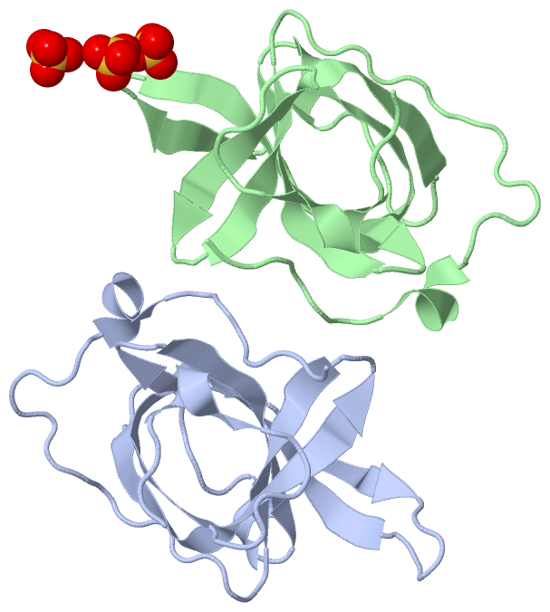
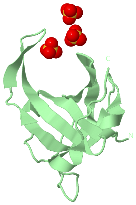
(-) Ligands, Modified Residues, Ions  (1, 3)

Asymmetric Unit (1, 3)
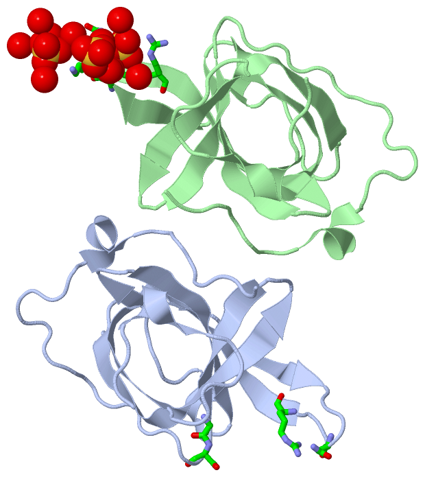
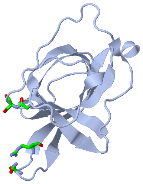
No.NameCountTypeFull Name
1SO43Ligand/IonSULFATE ION
Biological Unit 1 (0, 0)
No.NameCountTypeFull Name
1SO4-1Ligand/IonSULFATE ION
Biological Unit 2 (1, 3)
No.NameCountTypeFull Name
1SO43Ligand/IonSULFATE ION

(-) Sites  (3, 3)

Asymmetric Unit (3, 3)
No.NameEvidenceResiduesDescription
1AC1SOFTWAREASN A:34 , ARG A:37 , LYS B:33 , SO4 B:1116 , HOH B:2131 , HOH B:2135 , HOH B:2140BINDING SITE FOR RESIDUE SO4 B1115
2AC2SOFTWAREGLY A:88 , SER A:89 , LYS B:33 , SO4 B:1115 , HOH B:2132 , HOH B:2133 , HOH B:2134 , HOH B:2135 , HOH B:2136BINDING SITE FOR RESIDUE SO4 B1116
3AC3SOFTWAREASN A:34 , THR B:32 , ASN B:34 , ARG B:37 , HOH B:2137 , HOH B:2138 , HOH B:2140 , HOH B:2141BINDING SITE FOR RESIDUE SO4 B1117

(-) SS Bonds  (0, 0)

(no "SS Bond" information available for 1O7I)

(-) Cis Peptide Bonds  (0, 0)

(no "Cis Peptide Bond" information available for 1O7I)

 Sequence-Structure Mapping

(-) SAPs(SNPs)/Variants  (0, 0)

(no "SAP(SNP)/Variant" information available for 1O7I)

(-) PROSITE Motifs  (0, 0)

(no "PROSITE Motif" information available for 1O7I)

(-) Exons   (0, 0)

(no "Exon" information available for 1O7I)

(-) Sequences/Alignments

Asymmetric Unit
   Reformat: Number of residues per line =  ('0' or empty: single-line sequence representation)
  Number of residues per labelling interval =   
  UniProt sequence: complete  aligned part    
   Show mapping: SCOP domains CATH domains Pfam domains Secondary structure (by author)
SAPs(SNPs) PROSITE motifs Exons
(details for a mapped element are shown in a popup box when the mouse pointer rests over it)
Chain A from PDB  Type:PROTEIN  Length:115
 aligned with SSB_SULSO | Q97W73 from UniProtKB/Swiss-Prot  Length:148

    Alignment length:115
                                    10        20        30        40        50        60        70        80        90       100       110     
            SSB_SULSO     1 MEEKVGNLKPNMESVNVTVRVLEASEARQIQTKNGVRTISEAIVGDETGRVKLTLWGKHAGSIKEGQVVKIENAWTTAFKGQVQLNAGSKTKIAEASEDGFPESSQIPENTPTAP 115
               SCOP domains d1o7ia_ A: Archaeal ssDNA-binding protein                                                                           SCOP domains
               CATH domains 1o7iA00 A:1-115 Nucleic acid-binding proteins                                                                       CATH domains
               Pfam domains ------------------------------------------------------------------------------------------------------------------- Pfam domains
         Sec.struct. author ...hhhhh......eeeeeeeeee...eee......eeeeeeeeee..eeeeeeee.hhh.......eeeeeeeeeeee..eeeeee...............hhhhh........ Sec.struct. author
                 SAPs(SNPs) ------------------------------------------------------------------------------------------------------------------- SAPs(SNPs)
                    PROSITE ------------------------------------------------------------------------------------------------------------------- PROSITE
                 Transcript ------------------------------------------------------------------------------------------------------------------- Transcript
                 1o7i A   1 MEEKVGNLKPNMESVNVTVRVLEASEARQIQTKNGVRTISEAIVGDETGRVKLTLWGKHAGSIKEGQVVKIENAWTTAFKGQVQLNAGSKTKIAEASEDGFPESSQIPENTPTAP 115
                                    10        20        30        40        50        60        70        80        90       100       110     

Chain B from PDB  Type:PROTEIN  Length:114
 aligned with SSB_SULSO | Q97W73 from UniProtKB/Swiss-Prot  Length:148

    Alignment length:114
                                    10        20        30        40        50        60        70        80        90       100       110    
            SSB_SULSO     1 MEEKVGNLKPNMESVNVTVRVLEASEARQIQTKNGVRTISEAIVGDETGRVKLTLWGKHAGSIKEGQVVKIENAWTTAFKGQVQLNAGSKTKIAEASEDGFPESSQIPENTPTA 114
               SCOP domains d1o7ib_ B: Archaeal ssDNA-binding protein                                                                          SCOP domains
               CATH domains 1o7iB00 B:1-114 Nucleic acid-binding proteins                                                                      CATH domains
               Pfam domains ------------------------------------------------------------------------------------------------------------------ Pfam domains
         Sec.struct. author ...hhhhh......eeeeeeeeee...eeee....eeeeeeeeeee..eeeeeeee.hhh.......eeeeeeeeeeee..eeeeee...............hhhhh....... Sec.struct. author
                 SAPs(SNPs) ------------------------------------------------------------------------------------------------------------------ SAPs(SNPs)
                    PROSITE ------------------------------------------------------------------------------------------------------------------ PROSITE
                 Transcript ------------------------------------------------------------------------------------------------------------------ Transcript
                 1o7i B   1 MEEKVGNLKPNMESVNVTVRVLEASEARQIQTKNGVRTISEAIVGDETGRVKLTLWGKHAGSIKEGQVVKIENAWTTAFKGQVQLNAGSKTKIAEASEDGFPESSQIPENTPTA 114
                                    10        20        30        40        50        60        70        80        90       100       110    
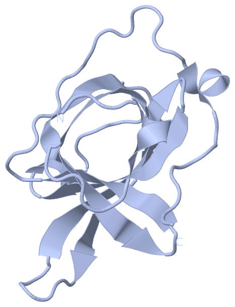
   Legend:   → Mismatch (orange background)
  - → Gap (green background, '-', border residues have a numbering label)
    → Modified Residue (blue background, lower-case, 'x' indicates undefined single-letter code, labelled with number + name)
  x → Chemical Group (purple background, 'x', labelled with number + name, e.g. ACE or NH2)
  extra numbering lines below/above indicate numbering irregularities and modified residue names etc., number ends below/above '|'

 Classification and Annotation

(-) SCOP Domains  (1, 2)

Asymmetric Unit

(-) CATH Domains  (1, 2)

Asymmetric Unit
(-)
Class: Mainly Beta (13760)

(-) Pfam Domains  (0, 0)

(no "Pfam Domain" information available for 1O7I)

(-) Gene Ontology  (2, 2)

Asymmetric Unit(hide GO term definitions)
Chain A,B   (SSB_SULSO | Q97W73)
molecular function
    GO:0003677    DNA binding    Any molecular function by which a gene product interacts selectively and non-covalently with DNA (deoxyribonucleic acid).
    GO:0003676    nucleic acid binding    Interacting selectively and non-covalently with any nucleic acid.

 Visualization

(-) Interactive Views

Asymmetric Unit
  Complete Structure
    Jena3D(integrated viewing of ligand, site, SAP, PROSITE, SCOP information)
    WebMol | AstexViewer[tm]@PDBe
(Java Applets, require no local installation except for Java; loading may be slow)
    STRAP
(Java WebStart application, automatic local installation, requires Java; full application with system access!)
    RasMol
(require local installation)
    Molscript (VRML)
(requires installation of a VRML viewer; select preferred view via VRML and generate a mono or stereo PDF format file)
 
  Ligands, Modified Residues, Ions
    SO4  [ RasMol | Jena3D ]  +environment [ RasMol | Jena3D ]
 
  Sites
    AC1  [ RasMol ]  +environment [ RasMol ]
    AC2  [ RasMol ]  +environment [ RasMol ]
    AC3  [ RasMol ]  +environment [ RasMol ]
 
  Cis Peptide Bonds
(no "Cis Peptide Bonds" information available for 1o7i)
 
Biological Units
  Complete Structure
    Biological Unit 1  [ Jena3D ]
    Biological Unit 2  [ Jena3D ]

(-) Still Images

Jmol
  protein: cartoon or spacefill or dots and stick; nucleic acid: cartoon and stick; ligands: spacefill; active site: stick
Molscript
  protein, nucleic acid: cartoon; ligands: spacefill; active site: ball and stick

 Databases and Analysis Tools

(-) Databases

Access by PDB/NDB ID
  1o7i
    Family and Domain InformationProDom | SYSTERS
    General Structural InformationGlycoscienceDB | MMDB | NDB | OCA | PDB | PDBe | PDBj | PDBsum | PDBWiki | PQS | PROTEOPEDIA
    Orientation in MembranesOPM
    Protein SurfaceSURFACE
    Secondary StructureDSSP (structure derived) | HSSP (homology derived)
    Structural GenomicsGeneCensus
    Structural NeighboursCE | VAST
    Structure ClassificationCATH | Dali | SCOP
    Validation and Original DataBMRB Data View | BMRB Restraints Grid | EDS | PROCHECK | RECOORD | WHAT_CHECK
 
Access by UniProt ID/Accession number
  SSB_SULSO | Q97W73
    Comparative Protein Structure ModelsModBase
    Genomic InformationEnsembl
    Protein-protein InteractionDIP
    Sequence, Family and Domain InformationInterPro | Pfam | SMART | UniProtKB/SwissProt
 
Access by Enzyme Classificator   (EC Number)
  (no 'Enzyme Classificator' available)
    General Enzyme InformationBRENDA | EC-PDB | Enzyme | IntEnz
    PathwayKEGG | MetaCyc
 
Access by Disease Identifier   (MIM ID)
  (no 'MIM ID' available)
    Disease InformationOMIM
 
Access by GenAge ID
  (no 'GenAge ID' available)
    Age Related InformationGenAge

(-) Analysis Tools

Access by PDB/NDB ID
    Domain InformationXDom
    Interatomic Contacts of Structural UnitsCSU
    Ligand-protein ContactsLPC
    Protein CavitiescastP
    Sequence and Secondary StructurePDBCartoon
    Structure AlignmentSTRAP(Java WebStart application, automatic local installation, requires Java; full application with system access!)
    Structure and Sequence BrowserSTING
 
Access by UniProt ID/Accession number
  SSB_SULSO | Q97W73
    Protein Disorder PredictionDisEMBL | FoldIndex | GLOBPLOT (for more information see DisProt)

 Related Entries

(-) Entries Sharing at Least One Protein Chain (UniProt ID)

UniProtKB/Swiss-Prot
        SSB_SULSO | Q97W732mna

(-) Related Entries Specified in the PDB File

(no "Related Entries Specified in the PDB File" available for 1O7I)