|
|
|
|
 Description
|
|
Compounds
|
||||||||||||||||||||||||||||||||||||||||
Chains, Units
Summary Information (see also Sequences/Alignments below) |
Ligands, Modified Residues, Ions (5, 7)
Asymmetric Unit (5, 7)
|
Sites (4, 4)
Asymmetric Unit (4, 4)
|
SS Bonds (0, 0)| (no "SS Bond" information available for 1O6B) |
Cis Peptide Bonds (1, 1)
Asymmetric Unit
|
||||||||
SAPs(SNPs)/Variants (0, 0)| (no "SAP(SNP)/Variant" information available for 1O6B) |
PROSITE Motifs (0, 0)| (no "PROSITE Motif" information available for 1O6B) |
Exons (0, 0)| (no "Exon" information available for 1O6B) |
Sequences/Alignments
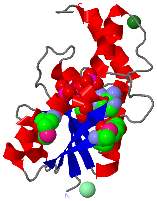
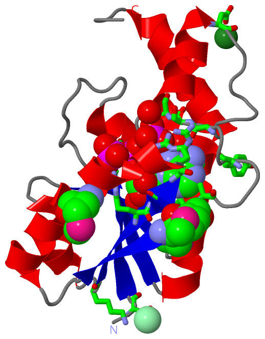
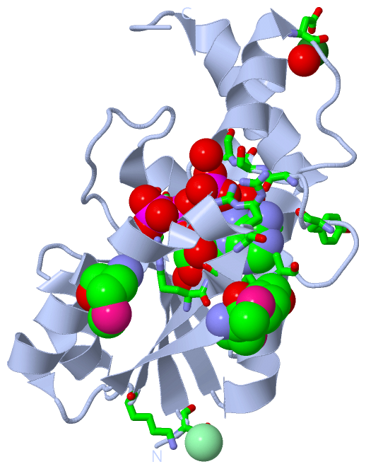
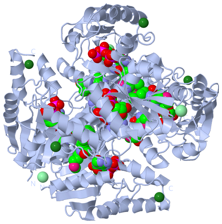
Asymmetric UnitChain A from PDB Type:PROTEIN Length:163 aligned with COAD_BACSU | O34797 from UniProtKB/Swiss-Prot Length:161 Alignment length:163 161 11 21 31 41 51 61 71 81 91 101 111 121 131 141 151 161 COAD_BACSU 2 ASIAVCPGSFDPVTYGHLDIIKRGAHIFEQVYVCVLNNSSKKPLFSVEERCELLREVTKDIPNITVETSQGLLIDYAKRKNAKAILRGLRAVSDFEYEMQGTSVNRVLDESIETFFMMTNNQYSFLSSSIVKEVARYNGSVSEFVPPEVELALQQKFRQG--- - SCOP domains d1o6ba_ A: Phosphopantetheine adenylyltransferase SCOP domains CATH domains 1o6bA00 A:2-164 Tyrosyl-Transfer RNA Synthetase , subunit E, domain 1 CATH domains Pfam domains ----CTP_transf_2-1o6bA01 A:6-135 ----------------------------- Pfam domains SAPs(SNPs) ------------------------------------------------------------------------------------------------------------------------------------------------------------------- SAPs(SNPs) PROSITE ------------------------------------------------------------------------------------------------------------------------------------------------------------------- PROSITE Transcript ------------------------------------------------------------------------------------------------------------------------------------------------------------------- Transcript 1o6b A 2 ASIAVCPGSFDPVTYGHLDIIKRGAHIFEQVYVCVLNNSSKKPLFSVEERCELLREVTKDIPNITVETSQGLLIDYARRKNAKAILRGLRAVSDFEYEmQGTSVNRVLDESIETFFmmANNQYSFLSSSIVKEVARYDGSVSEFVPPEVELALQQKFRQGGSH 164 11 21 31 41 51 61 71 81 91 101 111 |121 131 141 151 161 100-MSE 118-MSE 119-MSE
|
||||||||||||||||||||
SCOP Domains (1, 1)
Asymmetric Unit
 |
CATH Domains (1, 1)
Asymmetric Unit
|
Pfam Domains (1, 1)
Asymmetric Unit
|
Gene Ontology (9, 9)|
Asymmetric Unit(hide GO term definitions) Chain A (COAD_BACSU | O34797)
|
||||||||||||||||||||||||||||||||||||||||||||||||||||||||||||||||||||||||
Interactive Views
|
|||||||||||||||||||||||||||||||||||||||||||||||||||||||||||||||||||||||||||||||||||||||||||||||||||||||||||||||||||||||||||||||||||||||||||||||||||||||||||||||||||||||||||||||||||||||||||||||
Still Images
|
||||||||||||||||
Databases
|
||||||||||||||||||||||||||||||||||||||||||||||||||||||||||||||||||||||||||||||||||||||||||||||||||||||||||||||||||||||||||||||||||||||||||||||||||||||||||||||||
Analysis Tools
|
|||||||||||||||||||||||||||||||||||||||||||||||||||||||||||||
Entries Sharing at Least One Protein Chain (UniProt ID)
Related Entries Specified in the PDB File
|
|