Show PDB file:   
         Plain Text   HTML   (compressed file size)
QuickSearch:   
by PDB,NDB,UniProt,PROSITE Code or Search Term(s)  
(-)NMR Structure - model 1
(-)NMR Structure - model 1, sites
(-)NMR Structure - all models
collapse expand < >
Image NMR Structure - model 1
NMR Structure - model 1  (Jmol Viewer)
Image NMR Structure - model 1, sites
NMR Structure - model 1, sites  (Jmol Viewer)
Image NMR Structure - all models
NMR Structure - all models  (Jmol Viewer)

(-) Description

Title :  NMR SOLUTION STRUCTURE OF CALERYTHRIN, AN EF-HAND CALCIUM-BINDING PROTEIN
 
Authors :  H. Tossavainen, P. Permi, A. Annila, I. Kilpelainen, T. Drakenberg
Date :  12 Feb 03  (Deposition) - 05 Aug 03  (Release) - 24 Feb 09  (Revision)
Method :  SOLUTION NMR
Resolution :  NOT APPLICABLE
Chains :  NMR Structure  :  A  (20x)
Keywords :  Ef-Hand, Metal Binding Protein (Keyword Search: [Gene Ontology, PubMed, Web (Google))
 
Reference :  H. Tossavainen, P. Permi, A. Annila, I. Kilpelainen, T. Drakenberg
Nmr Solution Structure Of Calerythrin, An Ef-Hand Calcium-Binding Protein From Saccharopolyspora Erythraea
Eur. J. Biochem. V. 270 2505 2003
PubMed-ID: 12755706  |  Reference-DOI: 10.1046/J.1432-1033.2003.03623.X
(for further references see the PDB file header)

(-) Compounds

Molecule 1 - CALERYTHRIN
    ChainsA
    EngineeredYES
    Expression SystemESCHERICHIA COLI
    Expression System Taxid562
    MutationYES
    Organism ScientificSACCHAROPOLYSPORA ERYTHRAEA
    Organism Taxid1836
    SynonymCALCIUM-BINDING PROTEIN

 Structural Features

(-) Chains, Units

  
NMR Structure (20x)

Summary Information (see also Sequences/Alignments below)

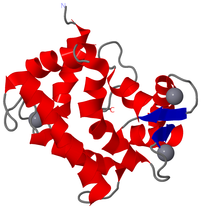
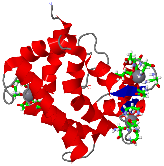
(-) Ligands, Modified Residues, Ions  (1, 3)

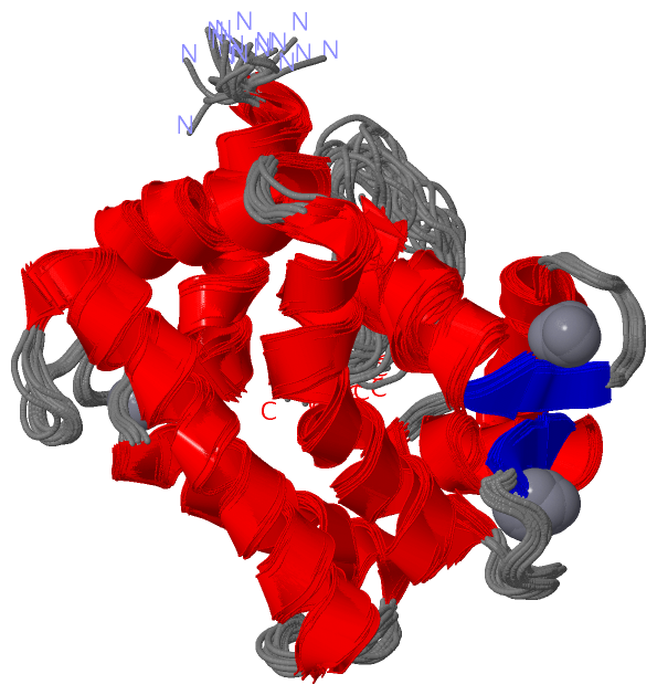
NMR Structure (1, 3)
No.NameCountTypeFull Name
1CA3Ligand/IonCALCIUM ION

(-) Sites  (3, 3)

NMR Structure (3, 3)
No.NameEvidenceResiduesDescription
1AC1SOFTWAREASP A:17 , PHE A:18 , ASP A:19 , ASN A:21 , ALA A:23 , ASP A:28BINDING SITE FOR RESIDUE CA A 500
2AC2SOFTWAREASP A:112 , ASN A:114 , ASP A:116 , GLN A:118 , GLU A:123BINDING SITE FOR RESIDUE CA A 501
3AC3SOFTWAREASP A:146 , THR A:147 , ASN A:148 , ASN A:150 , GLU A:152 , GLU A:157BINDING SITE FOR RESIDUE CA A 502

(-) SS Bonds  (0, 0)

(no "SS Bond" information available for 1NYA)

(-) Cis Peptide Bonds  (0, 0)

(no "Cis Peptide Bond" information available for 1NYA)

 Sequence-Structure Mapping

(-) SAPs(SNPs)/Variants  (0, 0)

(no "SAP(SNP)/Variant" information available for 1NYA)

(-) PROSITE Motifs  (2, 6)

NMR Structure (2, 6)
 PROSITEUniProtKBPDB
No.IDACDescriptionIDLocationCountLocation
1EF_HAND_2PS50222 EF-hand calcium-binding domain profile.CBP_SACER5-40
134-169
100-133
  3A:4-39
A:133-168
A:99-132
2EF_HAND_1PS00018 EF-hand calcium-binding domain.CBP_SACER18-30
113-125
147-159
  3A:17-29
A:112-124
A:146-158

(-) Exons   (0, 0)

(no "Exon" information available for 1NYA)

(-) Sequences/Alignments

NMR Structure
   Reformat: Number of residues per line =  ('0' or empty: single-line sequence representation)
  Number of residues per labelling interval =   
  UniProt sequence: complete  aligned part    
   Show mapping: SCOP domains CATH domains Pfam domains Secondary structure (by author)
SAPs(SNPs) PROSITE motifs Exons
(details for a mapped element are shown in a popup box when the mouse pointer rests over it)
Chain A from PDB  Type:PROTEIN  Length:176
 aligned with CBP_SACER | P06495 from UniProtKB/Swiss-Prot  Length:177

    Alignment length:176
                                    11        21        31        41        51        61        71        81        91       101       111       121       131       141       151       161       171      
            CBP_SACER     2 TTAIASDRLKKRFDRWDFDGNGALERADFEKEAQHIAEAFGKDAGAAEVQTLKNAFGGLFDYLAKEAGVGSDGSLTEEQFIRVTENLIFEQGEASFNRVLGPVVKGTWGMCDKNADGQINADEFAAWLTALGMSKAEAAEAFNQVDTNGNGELSLDELLTAVRDFHFGRLDVELLG 177
               SCOP domains d1nyaa_ A: Calerythrin                                                                                                                                                           SCOP domains
               CATH domains 1nyaA00 A:1-176 EF-hand                                                                                                                                                          CATH domains
               Pfam domains ----------------------------------------------------------------------------------------------------------------------------------------efhand-1nyaA01 A:137-165     ----------- Pfam domains
         Sec.struct. author ..hhhhhhhhhhhhhh.........hhhhhhhhhhhhhhh.....hhhhhhhhhhhhhhhhhhhhhhh.......hhhhhhhhhhhhhh..hhhhhhhhhhhhhhhhhhhh......eeehhhhhhhhhhh..hhhhhhhhhhhhh.....eeehhhhhhhhh............. Sec.struct. author
                 SAPs(SNPs) -------------------------------------------------------------------------------------------------------------------------------------------------------------------------------- SAPs(SNPs)
                PROSITE (1) ---EF_HAND_2  PDB: A:4-39 UniProt: 5-40-----------------------------------------------------------EF_HAND_2  PDB: A:99-132          EF_HAND_2  PDB: A:133-168           -------- PROSITE (1)
                PROSITE (2) ----------------EF_HAND_1    ----------------------------------------------------------------------------------EF_HAND_1    ---------------------EF_HAND_1    ------------------ PROSITE (2)
                 Transcript -------------------------------------------------------------------------------------------------------------------------------------------------------------------------------- Transcript
                 1nya A   1 TTAIASDRLKKRFDRWDFDGNGALERADFEKEAQHIAEAFGKDAGAAEVQTLKNAFGGLFDYLAKEAGVGSDGSLTEEQFIRVTENLIFEQGEASFNRVLGPVVKGIVGMCDKNADGQINADEFAAWLTALGMSKAEAAEAFNQVDTNGNGELSLDELLTAVRDFHFGRLDVELLG 176
                                    10        20        30        40        50        60        70        80        90       100       110       120       130       140       150       160       170      

   Legend:   → Mismatch (orange background)
  - → Gap (green background, '-', border residues have a numbering label)
    → Modified Residue (blue background, lower-case, 'x' indicates undefined single-letter code, labelled with number + name)
  x → Chemical Group (purple background, 'x', labelled with number + name, e.g. ACE or NH2)
  extra numbering lines below/above indicate numbering irregularities and modified residue names etc., number ends below/above '|'

 Classification and Annotation

(-) SCOP Domains  (1, 1)

NMR Structure

(-) CATH Domains  (1, 1)

NMR Structure

(-) Pfam Domains  (1, 1)

NMR Structure
(-)
Clan: EF_hand (270)

(-) Gene Ontology  (2, 2)

NMR Structure(hide GO term definitions)
Chain A   (CBP_SACER | P06495)
molecular function
    GO:0005509    calcium ion binding    Interacting selectively and non-covalently with calcium ions (Ca2+).
    GO:0046872    metal ion binding    Interacting selectively and non-covalently with any metal ion.

 Visualization

(-) Interactive Views

NMR Structure
  Complete Structure
    Jena3D(integrated viewing of ligand, site, SAP, PROSITE, SCOP information)
    WebMol | AstexViewer[tm]@PDBe
(Java Applets, require no local installation except for Java; loading may be slow)
    STRAP
(Java WebStart application, automatic local installation, requires Java; full application with system access!)
    RasMol
(require local installation)
    Molscript (VRML)
(requires installation of a VRML viewer; select preferred view via VRML and generate a mono or stereo PDF format file)
 
  Ligands, Modified Residues, Ions
    CA  [ RasMol | Jena3D ]  +environment [ RasMol | Jena3D ]
 
  Sites
    AC1  [ RasMol ]  +environment [ RasMol ]
    AC2  [ RasMol ]  +environment [ RasMol ]
    AC3  [ RasMol ]  +environment [ RasMol ]
 
  Cis Peptide Bonds
(no "Cis Peptide Bonds" information available for 1nya)
 

(-) Still Images

Jmol
  protein: cartoon or spacefill or dots and stick; nucleic acid: cartoon and stick; ligands: spacefill; active site: stick
Molscript
  protein, nucleic acid: cartoon; ligands: spacefill; active site: ball and stick

 Databases and Analysis Tools

(-) Databases

Access by PDB/NDB ID
  1nya
    Family and Domain InformationProDom | SYSTERS
    General Structural InformationGlycoscienceDB | MMDB | NDB | OCA | PDB | PDBe | PDBj | PDBsum | PDBWiki | PQS | PROTEOPEDIA
    Orientation in MembranesOPM
    Protein SurfaceSURFACE
    Secondary StructureDSSP (structure derived) | HSSP (homology derived)
    Structural GenomicsGeneCensus
    Structural NeighboursCE | VAST
    Structure ClassificationCATH | Dali | SCOP
    Validation and Original DataBMRB Data View | BMRB Restraints Grid | EDS | PROCHECK | RECOORD | WHAT_CHECK
 
Access by UniProt ID/Accession number
  CBP_SACER | P06495
    Comparative Protein Structure ModelsModBase
    Genomic InformationEnsembl
    Protein-protein InteractionDIP
    Sequence, Family and Domain InformationInterPro | Pfam | SMART | UniProtKB/SwissProt
 
Access by Enzyme Classificator   (EC Number)
  (no 'Enzyme Classificator' available)
    General Enzyme InformationBRENDA | EC-PDB | Enzyme | IntEnz
    PathwayKEGG | MetaCyc
 
Access by Disease Identifier   (MIM ID)
  (no 'MIM ID' available)
    Disease InformationOMIM
 
Access by GenAge ID
  (no 'GenAge ID' available)
    Age Related InformationGenAge

(-) Analysis Tools

Access by PDB/NDB ID
    Domain InformationXDom
    Interatomic Contacts of Structural UnitsCSU
    Ligand-protein ContactsLPC
    Protein CavitiescastP
    Sequence and Secondary StructurePDBCartoon
    Structure AlignmentSTRAP(Java WebStart application, automatic local installation, requires Java; full application with system access!)
    Structure and Sequence BrowserSTING
 
Access by UniProt ID/Accession number
  CBP_SACER | P06495
    Protein Disorder PredictionDisEMBL | FoldIndex | GLOBPLOT (for more information see DisProt)

 Related Entries

(-) Entries Sharing at Least One Protein Chain (UniProt ID)

(no "Entries Sharing at Least One Protein Chain" available for 1NYA)

(-) Related Entries Specified in the PDB File

(no "Related Entries Specified in the PDB File" available for 1NYA)