Show PDB file:   
         Plain Text   HTML   (compressed file size)
QuickSearch:   
by PDB,NDB,UniProt,PROSITE Code or Search Term(s)  
(-)Asym./Biol. Unit
(-)Asym./Biol. Unit - sites
collapse expand < >
Image Asym./Biol. Unit
Asym./Biol. Unit  (Jmol Viewer)
Image Asym./Biol. Unit - sites
Asym./Biol. Unit - sites  (Jmol Viewer)

(-) Description

Title :  STRUCTURE OF MOUSE GOLGI ALPHA-1,2-MANNOSIDASE IA REVEALS THE MOLECULAR BASIS FOR SUBSTRATE SPECIFICITY AMONG CLASS I ENZYMES (FAMILY 47 GLYCOSIDASES)
 
Authors :  W. Tempel, Z. -J. Liu, K. Karaveg, J. Rose, K. W. Moremen, B. -C. Wang, Sou Collaboratory For Structural Genomics (Secsg)
Date :  10 Feb 03  (Deposition) - 04 May 04  (Release) - 13 Jul 11  (Revision)
Method :  X-RAY DIFFRACTION
Resolution :  1.51
Chains :  Asym./Biol. Unit :  A
Keywords :  Mannosidase, Glycosidase, Structural Genomics, Psi, Protein Structure Initiative, Southeast Collaboratory For Structural Genomics, Secsg, Hydrolase (Keyword Search: [Gene Ontology, PubMed, Web (Google))
 
Reference :  W. Tempel, K. Karaveg, Z. -J. Liu, J. Rose, B. -C. Wang, K. W. Moremen
Structure Of Mouse Golgi Alpha-Mannosidase Ia Reveals The Molecular Basis For Substrate Specificity Among Class 1 (Family 47 Glycosylhydrolase) Alpha1, 2-Mannosidases
J. Biol. Chem. V. 279 29774 2004
PubMed-ID: 15102839  |  Reference-DOI: 10.1074/JBC.M403065200

(-) Compounds

Molecule 1 - MANNOSYL-OLIGOSACCHARIDE 1,2-ALPHA-MANNOSIDASE IA
    ChainsA
    EC Number3.2.1.113
    EngineeredYES
    Expression SystemPICHIA PASTORIS
    Expression System PlasmidPHIL S1
    Expression System StrainGS115
    Expression System Taxid4922
    Expression System Vector TypePLASMID
    GeneMAN1A
    Organism CommonHOUSE MOUSE
    Organism ScientificMUS MUSCULUS
    Organism Taxid10090
    SynonymPROCESSING ALPHA-1,2-MANNOSIDASE IA, ALPHA-1,2-MANNOSIDASE IA, MANNOSIDASE ALPHA CLASS 1A MEMBER 1, MAN(9)-ALPHA-MANNOSIDASE

 Structural Features

(-) Chains, Units

  1
Asymmetric/Biological Unit A

Summary Information (see also Sequences/Alignments below)
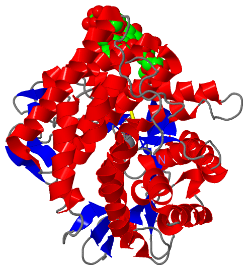
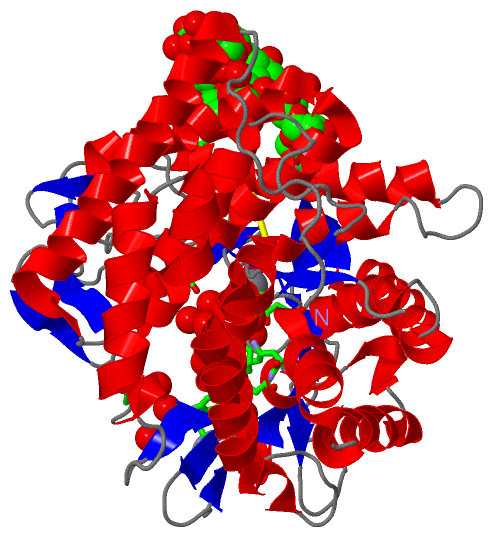
(-) Ligands, Modified Residues, Ions  (4, 9)

Asymmetric/Biological Unit (4, 9)
No.NameCountTypeFull Name
1BMA1Ligand/IonBETA-D-MANNOSE
2CA1Ligand/IonCALCIUM ION
3MAN5Ligand/IonALPHA-D-MANNOSE
4NAG2Ligand/IonN-ACETYL-D-GLUCOSAMINE

(-) Sites  (9, 9)

Asymmetric Unit (9, 9)
No.NameEvidenceResiduesDescription
1AC1SOFTWARENAG A:2 , TRP A:341 , GLU A:512 , ASN A:515 , HOH A:1060 , HOH A:1104 , HOH A:1227 , HOH A:1248 , HOH A:1294BINDING SITE FOR RESIDUE NAG A 1
2AC2SOFTWARENAG A:1 , BMA A:3 , MAN A:7 , TRP A:341 , LEU A:413 , HOH A:1045 , HOH A:1243 , HOH A:1322BINDING SITE FOR RESIDUE NAG A 2
3AC3SOFTWARENAG A:2 , MAN A:4 , MAN A:5 , HOH A:1117 , HOH A:1184BINDING SITE FOR RESIDUE BMA A 3
4AC4SOFTWAREBMA A:3 , MAN A:6 , MAN A:7 , LEU A:413 , GLU A:524 , HOH A:1045 , HOH A:1172 , HOH A:1262BINDING SITE FOR RESIDUE MAN A 4
5AC5SOFTWAREMAN A:4 , PHE A:281 , ALA A:350 , GLU A:351 , LEU A:413 , ASP A:415 , HOH A:1012 , HOH A:1036 , HOH A:1058 , HOH A:1079BINDING SITE FOR RESIDUE MAN A 6
6AC6SOFTWARENAG A:2 , MAN A:4 , HOH A:1126 , HOH A:1197 , HOH A:1243BINDING SITE FOR RESIDUE MAN A 7
7AC7SOFTWAREBMA A:3 , MAN A:8 , SER A:233 , HOH A:1270BINDING SITE FOR RESIDUE MAN A 5
8AC8SOFTWAREMAN A:5 , SER A:455 , GLY A:456 , HOH A:1139 , HOH A:1270BINDING SITE FOR RESIDUE MAN A 8
9AC9SOFTWARETHR A:635 , HOH A:1086 , HOH A:1087 , HOH A:1088 , HOH A:1089 , HOH A:1090 , HOH A:1091BINDING SITE FOR RESIDUE CA A 656

(-) SS Bonds  (1, 1)

Asymmetric/Biological Unit
No.Residues
1A:478 -A:510

(-) Cis Peptide Bonds  (0, 0)

(no "Cis Peptide Bond" information available for 1NXC)

 Sequence-Structure Mapping

(-) SAPs(SNPs)/Variants  (0, 0)

(no "SAP(SNP)/Variant" information available for 1NXC)

(-) PROSITE Motifs  (0, 0)

(no "PROSITE Motif" information available for 1NXC)

(-) Exons   (0, 0)

(no "Exon" information available for 1NXC)

(-) Sequences/Alignments

Asymmetric/Biological Unit
   Reformat: Number of residues per line =  ('0' or empty: single-line sequence representation)
  Number of residues per labelling interval =   
  UniProt sequence: complete  aligned part    
   Show mapping: SCOP domains CATH domains Pfam domains Secondary structure (by author)
SAPs(SNPs) PROSITE motifs Exons
(details for a mapped element are shown in a popup box when the mouse pointer rests over it)
Chain A from PDB  Type:PROTEIN  Length:467
 aligned with MA1A1_MOUSE | P45700 from UniProtKB/Swiss-Prot  Length:655

    Alignment length:467
                                   187       197       207       217       227       237       247       257       267       277       287       297       307       317       327       337       347       357       367       377       387       397       407       417       427       437       447       457       467       477       487       497       507       517       527       537       547       557       567       577       587       597       607       617       627       637       
          MA1A1_MOUSE   178 FLPPVGVENREPADATIREKRAKIKEMMTHAWNNYKRYAWGLNELKPISKEGHSSSLFGNIKGATIVDALDTLFIMGMKTEFQEAKSWIKKYLDFNVNAEVSVFEVNIRFVGGLLSAYYLSGEEIFRKKAVELGVKLLPAFHTPSGIPWALLNMKSGIGRNWPWASGGSSILAEFGTLHLEFMHLSHLSGDPVFAEKVMKIRTVLNKLDKPEGLYPNYLNPSSGQWGQHHVSVGGLGDSFYEYLLKAWLMSDKTDLEAKKMYFDAVQAIETHLIRKSSGGLTYIAEWKGGLLEHKMGHLTCFAGGMFALGADGAPEARAQHYLELGAEIARTCHESYNRTYVKLGPEAFRFDGGVEAIATRQNEKYYILRPEVIETYMYMWRLTHDPKYRTWAWEAVEALESHCRVNGGYSGLRDVYIARESYDDVQQSFFLAETLKYLYLIFSDDDLLPLEHWIFNTEAHPFPILR 644
               SCOP domains d1nxca_ A: Class I alpha-1;2-mannosidase, catalytic domain                                                                                                                                                                                                                                                                                                                                                                                                                          SCOP domains
               CATH domains 1nxcA00 A:178-644  [code=1.50.10.50, no name defined]                                                                                                                                                                                                                                                                                                                                                                                                                               CATH domains
               Pfam domains --------------------------Glyco_hydro_47-1nxcA01 A:204-642                                                                                                                                                                                                                                                                                                                                                                                                                       -- Pfam domains
         Sec.struct. author .............hhhhhhhhhhhhhhhhhhhhhhhhhhh...eee....eee.hhhhh...hhhhhhhhhhhhhh.hhhhhhhhhhhhhhhh......eeehhhhhhhhhhhhhhhhhhhhhhhhhhhhhhhhhhhhhhhh........eee.....ee....hhhhheehhhhh.hhhhhhhhhhhhh.hhhhhhhhhhhhhhhhh.hhhhh...ee..........ee.....hhhhhhhhhhhhhhh...hhhhhhhhhhhhhhhhhhheee.....eee.eee..eee.eee.hhhhhhhhhhhh.......hhhhhhhhhhhhhhhhhhhhhh........eee..........hhhhh.....hhhhhhhhhhhhhhhhhhhhhhhhhhhhhhhhhhee..ee.................hhhhhhhhhhhhhhhh..........eee.....eee... Sec.struct. author
                 SAPs(SNPs) ----------------------------------------------------------------------------------------------------------------------------------------------------------------------------------------------------------------------------------------------------------------------------------------------------------------------------------------------------------------------------------------------------------------------------------------------------------------------------------- SAPs(SNPs)
                    PROSITE ----------------------------------------------------------------------------------------------------------------------------------------------------------------------------------------------------------------------------------------------------------------------------------------------------------------------------------------------------------------------------------------------------------------------------------------------------------------------------------- PROSITE
                 Transcript ----------------------------------------------------------------------------------------------------------------------------------------------------------------------------------------------------------------------------------------------------------------------------------------------------------------------------------------------------------------------------------------------------------------------------------------------------------------------------------- Transcript
                 1nxc A 178 FLPPVGVENREPADATIREKRAKIKEMMTHAWNNYKRYAWGLNELKPISKEGHSSSLFGNIKGATIVDALDTLFIMGMKTEFQEAKSWIKKYLDFNVNAEVSVFEVNIRFVGGLLSAYYLSGEEIFRKKAVELGVKLLPAFHTPSGIPWALLNMKSGIGRNWPWASGGSSILAEFGTLHLEFMHLSHLSGDPVFAEKVMKIRTVLNKLDKPEGLYPNYLNPSSGQWGQHHVSVGGLGDSFYEYLLKAWLMSDKTDLEAKKMYFDAVQAIETHLIRKSSGGLTYIAEWKGGLLEHKMGHLTCFAGGMFALGADGAPEARAQHYLELGAEIARTCHESYNRTYVKLGPEAFRFDGGVEAIATRQNEKYYILRPEVIETYMYMWRLTHDPKYRTWAWEAVEALESHCRVNGGYSGLRDVYIARESYDDVQQSFFLAETLKYLYLIFSDDDLLPLEHWIFNTEAHPFPILR 644
                                   187       197       207       217       227       237       247       257       267       277       287       297       307       317       327       337       347       357       367       377       387       397       407       417       427       437       447       457       467       477       487       497       507       517       527       537       547       557       567       577       587       597       607       617       627       637       

   Legend:   → Mismatch (orange background)
  - → Gap (green background, '-', border residues have a numbering label)
    → Modified Residue (blue background, lower-case, 'x' indicates undefined single-letter code, labelled with number + name)
  x → Chemical Group (purple background, 'x', labelled with number + name, e.g. ACE or NH2)
  extra numbering lines below/above indicate numbering irregularities and modified residue names etc., number ends below/above '|'

 Classification and Annotation

(-) SCOP Domains  (1, 1)

Asymmetric/Biological Unit

(-) CATH Domains  (1, 1)

Asymmetric/Biological Unit

(-) Pfam Domains  (1, 1)

Asymmetric/Biological Unit
(-)
Clan: 6_Hairpin (120)

(-) Gene Ontology  (16, 16)

Asymmetric/Biological Unit(hide GO term definitions)
Chain A   (MA1A1_MOUSE | P45700)
molecular function
    GO:0005509    calcium ion binding    Interacting selectively and non-covalently with calcium ions (Ca2+).
    GO:0016787    hydrolase activity    Catalysis of the hydrolysis of various bonds, e.g. C-O, C-N, C-C, phosphoric anhydride bonds, etc. Hydrolase is the systematic name for any enzyme of EC class 3.
    GO:0016798    hydrolase activity, acting on glycosyl bonds    Catalysis of the hydrolysis of any glycosyl bond.
    GO:0004571    mannosyl-oligosaccharide 1,2-alpha-mannosidase activity    Catalysis of the hydrolysis of the terminal (1->2)-linked alpha-D-mannose residues in an oligo-mannose oligosaccharide.
    GO:0046872    metal ion binding    Interacting selectively and non-covalently with any metal ion.
biological process
    GO:0006491    N-glycan processing    The conversion of N-linked glycan (N = nitrogen) structures from the initially transferred oligosaccharide to a mature form, by the actions of glycosidases and glycosyltransferases. The early processing steps are conserved and play roles in glycoprotein folding and trafficking.
    GO:0008152    metabolic process    The chemical reactions and pathways, including anabolism and catabolism, by which living organisms transform chemical substances. Metabolic processes typically transform small molecules, but also include macromolecular processes such as DNA repair and replication, and protein synthesis and degradation.
    GO:0006486    protein glycosylation    A protein modification process that results in the addition of a carbohydrate or carbohydrate derivative unit to a protein amino acid, e.g. the addition of glycan chains to proteins.
cellular component
    GO:0005794    Golgi apparatus    A compound membranous cytoplasmic organelle of eukaryotic cells, consisting of flattened, ribosome-free vesicles arranged in a more or less regular stack. The Golgi apparatus differs from the endoplasmic reticulum in often having slightly thicker membranes, appearing in sections as a characteristic shallow semicircle so that the convex side (cis or entry face) abuts the endoplasmic reticulum, secretory vesicles emerging from the concave side (trans or exit face). In vertebrate cells there is usually one such organelle, while in invertebrates and plants, where they are known usually as dictyosomes, there may be several scattered in the cytoplasm. The Golgi apparatus processes proteins produced on the ribosomes of the rough endoplasmic reticulum; such processing includes modification of the core oligosaccharides of glycoproteins, and the sorting and packaging of proteins for transport to a variety of cellular locations. Three different regions of the Golgi are now recognized both in terms of structure and function: cis, in the vicinity of the cis face, trans, in the vicinity of the trans face, and medial, lying between the cis and trans regions.
    GO:0000139    Golgi membrane    The lipid bilayer surrounding any of the compartments of the Golgi apparatus.
    GO:0005737    cytoplasm    All of the contents of a cell excluding the plasma membrane and nucleus, but including other subcellular structures.
    GO:0005783    endoplasmic reticulum    The irregular network of unit membranes, visible only by electron microscopy, that occurs in the cytoplasm of many eukaryotic cells. The membranes form a complex meshwork of tubular channels, which are often expanded into slitlike cavities called cisternae. The ER takes two forms, rough (or granular), with ribosomes adhering to the outer surface, and smooth (with no ribosomes attached).
    GO:0005793    endoplasmic reticulum-Golgi intermediate compartment    A complex system of membrane-bounded compartments located between endoplasmic reticulum (ER) and the Golgi complex, with a distinctive membrane protein composition; involved in ER-to-Golgi and Golgi-to-ER transport.
    GO:0070062    extracellular exosome    A vesicle that is released into the extracellular region by fusion of the limiting endosomal membrane of a multivesicular body with the plasma membrane. Extracellular exosomes, also simply called exosomes, have a diameter of about 40-100 nm.
    GO:0016021    integral component of membrane    The component of a membrane consisting of the gene products and protein complexes having at least some part of their peptide sequence embedded in the hydrophobic region of the membrane.
    GO:0016020    membrane    A lipid bilayer along with all the proteins and protein complexes embedded in it an attached to it.

 Visualization

(-) Interactive Views

Asymmetric/Biological Unit
  Complete Structure
    Jena3D(integrated viewing of ligand, site, SAP, PROSITE, SCOP information)
    WebMol | AstexViewer[tm]@PDBe
(Java Applets, require no local installation except for Java; loading may be slow)
    STRAP
(Java WebStart application, automatic local installation, requires Java; full application with system access!)
    RasMol
(require local installation)
    Molscript (VRML)
(requires installation of a VRML viewer; select preferred view via VRML and generate a mono or stereo PDF format file)
 
  Ligands, Modified Residues, Ions
    BMA  [ RasMol | Jena3D ]  +environment [ RasMol | Jena3D ]
    CA  [ RasMol | Jena3D ]  +environment [ RasMol | Jena3D ]
    MAN  [ RasMol | Jena3D ]  +environment [ RasMol | Jena3D ]
    NAG  [ RasMol | Jena3D ]  +environment [ RasMol | Jena3D ]
 
  Sites
    AC1  [ RasMol ]  +environment [ RasMol ]
    AC2  [ RasMol ]  +environment [ RasMol ]
    AC3  [ RasMol ]  +environment [ RasMol ]
    AC4  [ RasMol ]  +environment [ RasMol ]
    AC5  [ RasMol ]  +environment [ RasMol ]
    AC6  [ RasMol ]  +environment [ RasMol ]
    AC7  [ RasMol ]  +environment [ RasMol ]
    AC8  [ RasMol ]  +environment [ RasMol ]
    AC9  [ RasMol ]  +environment [ RasMol ]
 
  Cis Peptide Bonds
(no "Cis Peptide Bonds" information available for 1nxc)
 

(-) Still Images

Jmol
  protein: cartoon or spacefill or dots and stick; nucleic acid: cartoon and stick; ligands: spacefill; active site: stick
Molscript
  protein, nucleic acid: cartoon; ligands: spacefill; active site: ball and stick

 Databases and Analysis Tools

(-) Databases

Access by PDB/NDB ID
  1nxc
    Family and Domain InformationProDom | SYSTERS
    General Structural InformationGlycoscienceDB | MMDB | NDB | OCA | PDB | PDBe | PDBj | PDBsum | PDBWiki | PQS | PROTEOPEDIA
    Orientation in MembranesOPM
    Protein SurfaceSURFACE
    Secondary StructureDSSP (structure derived) | HSSP (homology derived)
    Structural GenomicsGeneCensus
    Structural NeighboursCE | VAST
    Structure ClassificationCATH | Dali | SCOP
    Validation and Original DataBMRB Data View | BMRB Restraints Grid | EDS | PROCHECK | RECOORD | WHAT_CHECK
 
Access by UniProt ID/Accession number
  MA1A1_MOUSE | P45700
    Comparative Protein Structure ModelsModBase
    Genomic InformationEnsembl
    Protein-protein InteractionDIP
    Sequence, Family and Domain InformationInterPro | Pfam | SMART | UniProtKB/SwissProt
 
Access by Enzyme Classificator   (EC Number)
  3.2.1.113
    General Enzyme InformationBRENDA | EC-PDB | Enzyme | IntEnz
    PathwayKEGG | MetaCyc
 
Access by Disease Identifier   (MIM ID)
  (no 'MIM ID' available)
    Disease InformationOMIM
 
Access by GenAge ID
  (no 'GenAge ID' available)
    Age Related InformationGenAge

(-) Analysis Tools

Access by PDB/NDB ID
    Domain InformationXDom
    Interatomic Contacts of Structural UnitsCSU
    Ligand-protein ContactsLPC
    Protein CavitiescastP
    Sequence and Secondary StructurePDBCartoon
    Structure AlignmentSTRAP(Java WebStart application, automatic local installation, requires Java; full application with system access!)
    Structure and Sequence BrowserSTING
 
Access by UniProt ID/Accession number
  MA1A1_MOUSE | P45700
    Protein Disorder PredictionDisEMBL | FoldIndex | GLOBPLOT (for more information see DisProt)

 Related Entries

(-) Entries Sharing at Least One Protein Chain (UniProt ID)

UniProtKB/Swiss-Prot
        MA1A1_MOUSE | P457005kkb

(-) Related Entries Specified in the PDB File

(no "Related Entries Specified in the PDB File" available for 1NXC)