|
|
|
|
 Description
|
|
Compounds
|
||||||||||||||||||||||||||||||||||||||||||||||||
Chains, Units
Summary Information (see also Sequences/Alignments below) |
Ligands, Modified Residues, Ions (0, 0)| (no "Ligand,Modified Residues,Ions" information available for 1NSH) |
Sites (0, 0)| (no "Site" information available for 1NSH) |
SS Bonds (0, 0)| (no "SS Bond" information available for 1NSH) |
Cis Peptide Bonds (0, 0)| (no "Cis Peptide Bond" information available for 1NSH) |
SAPs(SNPs)/Variants (0, 0)| (no "SAP(SNP)/Variant" information available for 1NSH) |
PROSITE Motifs (3, 6)
NMR Structure (3, 6)
|
||||||||||||||||||||||||||||||||||||||||
Exons (0, 0)| (no "Exon" information available for 1NSH) |
Sequences/Alignments
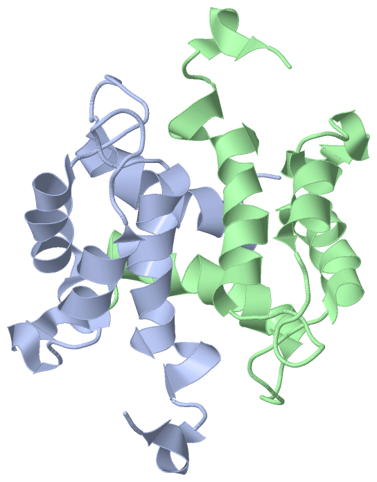
NMR StructureChain A from PDB Type:PROTEIN Length:101 aligned with S10AB_RABIT | P24480 from UniProtKB/Swiss-Prot Length:102 Alignment length:101 11 21 31 41 51 61 71 81 91 101 S10AB_RABIT 2 SRPTETERCIESLIAVFQKYAGKDGHSVTLSKTEFLSFMNTELAAFTKNQKDPGVLDRMMKKLDLNSDGQLDFQEFLNLIGGLAVACHESFVKAAPPQKRF 102 SCOP domains d1nsha_ A: Calcyclin (S100) SCOP domains CATH domains 1nshA00 A:1-101 EF-hand CATH domains Pfam domains ----------------------------------------------------------------------------------------------------- Pfam domains SAPs(SNPs) ----------------------------------------------------------------------------------------------------- SAPs(SNPs) PROSITE (1) --------------------------------------------------EF_HAND_2 PDB: A:51-86 --------------- PROSITE (1) PROSITE (2) ----------------------------------------------------------S100_CABP PDB: A:59-8--------------------- PROSITE (2) PROSITE (3) ---------------------------------------------------------------EF_HAND_1 ------------------------- PROSITE (3) Transcript ----------------------------------------------------------------------------------------------------- Transcript 1nsh A 1 SRPTETERCIESLIAVFQKYAGKDGHSVTLSKTEFLSFMNTELAAFTKNQKDPGVLDRMMKKLDLNSDGQLDFQEFLNLIGGLAVACHESFVKAAPPQKRF 101 10 20 30 40 50 60 70 80 90 100 Chain B from PDB Type:PROTEIN Length:101 aligned with S10AB_RABIT | P24480 from UniProtKB/Swiss-Prot Length:102 Alignment length:101 11 21 31 41 51 61 71 81 91 101 S10AB_RABIT 2 SRPTETERCIESLIAVFQKYAGKDGHSVTLSKTEFLSFMNTELAAFTKNQKDPGVLDRMMKKLDLNSDGQLDFQEFLNLIGGLAVACHESFVKAAPPQKRF 102 SCOP domains d1nshb_ B: Calcyclin (S100) SCOP domains CATH domains 1nshB00 B:1-101 EF-hand CATH domains Pfam domains (1) -----S_100-1nshB01 B:6-49 ---------------------------------------------------- Pfam domains (1) Pfam domains (2) -----S_100-1nshB02 B:6-49 ---------------------------------------------------- Pfam domains (2) SAPs(SNPs) ----------------------------------------------------------------------------------------------------- SAPs(SNPs) PROSITE (1) --------------------------------------------------EF_HAND_2 PDB: B:51-86 --------------- PROSITE (1) PROSITE (2) ----------------------------------------------------------S100_CABP PDB: B:59-8--------------------- PROSITE (2) PROSITE (3) ---------------------------------------------------------------EF_HAND_1 ------------------------- PROSITE (3) Transcript ----------------------------------------------------------------------------------------------------- Transcript 1nsh B 1 SRPTETERCIESLIAVFQKYAGKDGHSVTLSKTEFLSFMNTELAAFTKNQKDPGVLDRMMKKLDLNSDGQLDFQEFLNLIGGLAVACHESFVKAAPPQKRF 101 10 20 30 40 50 60 70 80 90 100
|
||||||||||||||||||||
SCOP Domains (1, 2)
NMR Structure
 |
CATH Domains (1, 2)
NMR Structure
|
Pfam Domains (1, 2)
NMR Structure
|
Gene Ontology (6, 6)|
NMR Structure(hide GO term definitions) Chain A,B (S10AB_RABIT | P24480)
|
||||||||||||||||||||||||||||||||||||||||||||||||||||||
Interactive Views
|
||||||||||||||||||||||||||||||||||||||||||||||||||||||||||||||||||||||||||||||||||||||||||||||||||||||||||||||||||||
Still Images
|
||||||||||||||||
Databases
|
||||||||||||||||||||||||||||||||||||||||||||||||||||||||||||||||||||||||||||||||||||||||||||||||||||||||||||||||||||||||||||||||||||||||||||||||||||||||||||||||
Analysis Tools
|
|||||||||||||||||||||||||||||||||||||||||||||||||||||||||||||
Entries Sharing at Least One Protein Chain (UniProt ID)
Related Entries Specified in the PDB File
|
|