|
|
|
|
 Description
|
|
Compounds
|
||||||||||||||||||||||||||||||||||||||||||||||||||||
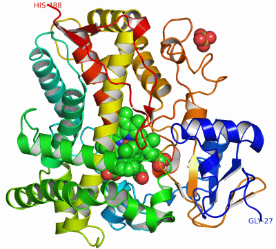
Chains, Units
Summary Information (see also Sequences/Alignments below) |
Ligands, Modified Residues, Ions (3, 4)| Asymmetric/Biological Unit (3, 4) |
Sites (4, 4)
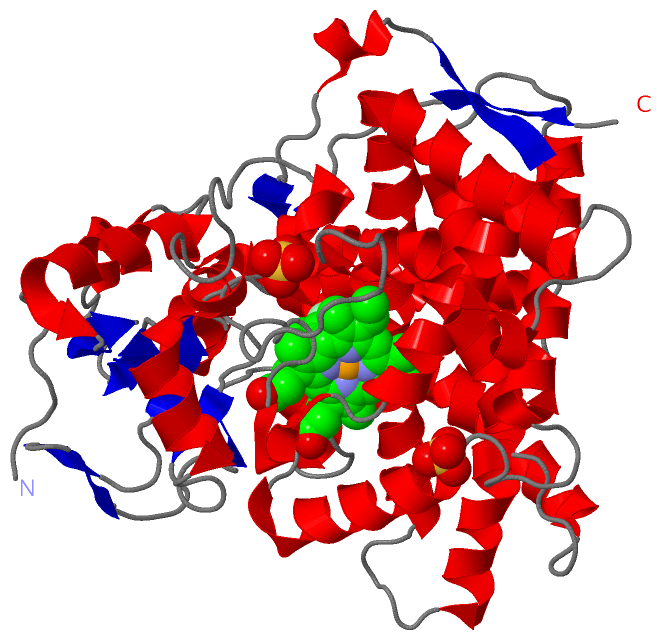
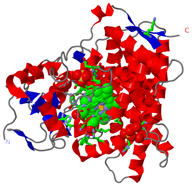
Asymmetric Unit (4, 4)
|
SS Bonds (0, 0)| (no "SS Bond" information available for 1NR6) |
Cis Peptide Bonds (0, 0)| (no "Cis Peptide Bond" information available for 1NR6) |
SAPs(SNPs)/Variants (0, 0)| (no "SAP(SNP)/Variant" information available for 1NR6) |
PROSITE Motifs (1, 1)
Asymmetric/Biological Unit (1, 1)
|
||||||||||||||||||||||||
Exons (0, 0)| (no "Exon" information available for 1NR6) |
Sequences/Alignments
Asymmetric/Biological UnitChain A from PDB Type:PROTEIN Length:462 aligned with CP2C5_RABIT | P00179 from UniProtKB/Swiss-Prot Length:487 Alignment length:462 487 36 46 56 66 76 86 96 106 116 126 136 146 156 166 176 186 196 206 216 226 236 246 256 266 276 286 296 306 316 326 336 346 356 366 376 386 396 406 416 426 436 446 456 466 476 486| CP2C5_RABIT 27 GKLPPGPTPFPIIGNILQIDAKDISKSLTKFSECYGPVFTVYLGMKPTVVLHGYEAVKEALVDLGEEFAGRGSVPILEKVSKGLGIAFSNAKTWKEMRRFSLMTLRNFGMGKRSIEDRIQEEARCLVEELRKTNASPCDPTFILGCAPCNVICSVIFHNRFDYKDEEFLKLMESLNENVRILSSPWLQVYNNFPALLDYFPGIHKTLLKNADYIKNFIMEKVKEHQKLLDVNNPRDFIDCFLIKMEQENNLEFTLESLVIAVSDLFGAGTETTSTTLRYSLLLLLKHPEVAARVQEEIERVIGRHRSPCMQDRSRMPYTDAVIHEIQRFIDLLPTNLPHAVTRDVRFRNYFIPKGTDIITSLTSVLHDEKAFPNPKVFDPGHFLDESGNFKKSDYFMPFSAGKRMCVGEGLARMELFLFLTSILQNFKLQSLVEPKDLDITAVVNGFVSVPPSYQLCFIPI- - SCOP domains d1nr6a_ A: Mammalian cytochrome p450 2c5 SCOP domains CATH domains 1nr6A00 A:27-488 Cytochrome p450 CATH domains Pfam domains ---p450-1nr6A01 A:30-484 ---- Pfam domains SAPs(SNPs) ------------------------------------------------------------------------------------------------------------------------------------------------------------------------------------------------------------------------------------------------------------------------------------------------------------------------------------------------------------------------------------------------------------------------------------------------------------------------------ SAPs(SNPs) PROSITE --------------------------------------------------------------------------------------------------------------------------------------------------------------------------------------------------------------------------------------------------------------------------------------------------------------------------------------------------------------------------------------------------------------CYTOCHROME------------------------------------------------------ PROSITE Transcript ------------------------------------------------------------------------------------------------------------------------------------------------------------------------------------------------------------------------------------------------------------------------------------------------------------------------------------------------------------------------------------------------------------------------------------------------------------------------------ Transcript 1nr6 A 27 GKLPPGPTPFPIIGNILQIDAKDISKSLTKFSECYGPVFTVYLGMKPTVVLHGYEAVKEALVDLGEEFAGRGSVPILEKVSKGLGIAFSNAKTWKEMRRFSLMTLRNFGMGKRSIEDRIQEEARCLVEELRKTNASPCDPTFILGCAPCNVICSVIFHNRFDYKDEEFLKLMESLHENVELLGTPWLQVYNNFPALLDYFPGIHKTLLKNADYIKNFIMEKVKEHQKLLDVNNPRDFIDCFLIKMEQENNLEFTLESLVIAVSDLFGAGTETTSTTLRYSLLLLLKHPEVAARVQEEIERVIGRHRSPCMQDRSRMPYTDAVIHEIQRFIDLLPTNLPHAVTRDVRFRNYFIPKGTDIITSLTSVLHDEKAFPNPKVFDPGHFLDESGNFKKSDYFMPFSAGKRMCVGEGLARMELFLFLTSILQNFKLQSLVEPKDLDITAVVNGFVSVPPSYQLCFIPIH 488 36 46 56 66 76 86 96 106 116 126 136 146 156 166 176 186 196 206 216 226 236 246 256 266 276 286 296 306 316 326 336 346 356 366 376 386 396 406 416 426 436 446 456 466 476 486
|
||||||||||||||||||||
SCOP Domains (1, 1)
Asymmetric/Biological Unit
 |
CATH Domains (1, 1)
Asymmetric/Biological Unit
|
Pfam Domains (1, 1)
Asymmetric/Biological Unit
|
Gene Ontology (13, 13)|
Asymmetric/Biological Unit(hide GO term definitions) Chain A (CP2C5_RABIT | P00179)
|
||||||||||||||||||||||||||||||||||||||||||||||||||||||||||||||||||||||||||||||||||||||||||||||||
Interactive Views
|
|||||||||||||||||||||||||||||||||||||||||||||||||||||||||||||||||||||||||||||||||||||||||||||||||||||||||||||||||||||||||||||||||||||||||||||||||||||||||
Still Images
|
||||||||||||||||
Databases
|
||||||||||||||||||||||||||||||||||||||||||||||||||||||||||||||||||||||||||||||||||||||||||||||||||||||||||||||||||||||||||||||||||||||||||||||||||||||||||||||||
Analysis Tools
|
|||||||||||||||||||||||||||||||||||||||||||||||||||||||||||||
Entries Sharing at Least One Protein Chain (UniProt ID)
Related Entries Specified in the PDB File
|
|