|
|
|
|
 Description
|
|
Compounds
|
||||||||||||||||||||||||
Chains, Units
Summary Information (see also Sequences/Alignments below) |
Ligands, Modified Residues, Ions (1, 2)
Asymmetric Unit (1, 2)
|
Sites (2, 2)
Asymmetric Unit (2, 2)
|
SS Bonds (0, 0)| (no "SS Bond" information available for 1MXS) |
Cis Peptide Bonds (1, 1)
Asymmetric Unit
|
||||||||
SAPs(SNPs)/Variants (0, 0)| (no "SAP(SNP)/Variant" information available for 1MXS) |
PROSITE Motifs (2, 2)
Asymmetric Unit (2, 2)
|
||||||||||||||||||||||||||||||||||||||||||||||||||||||||||||||||
Exons (0, 0)| (no "Exon" information available for 1MXS) |
Sequences/Alignments
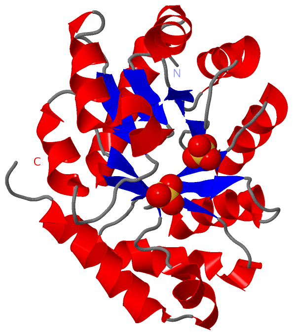
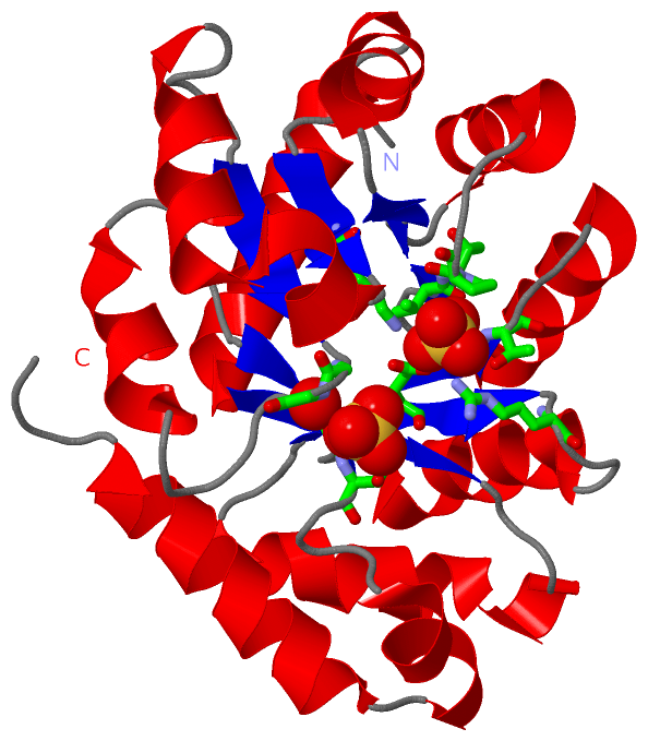
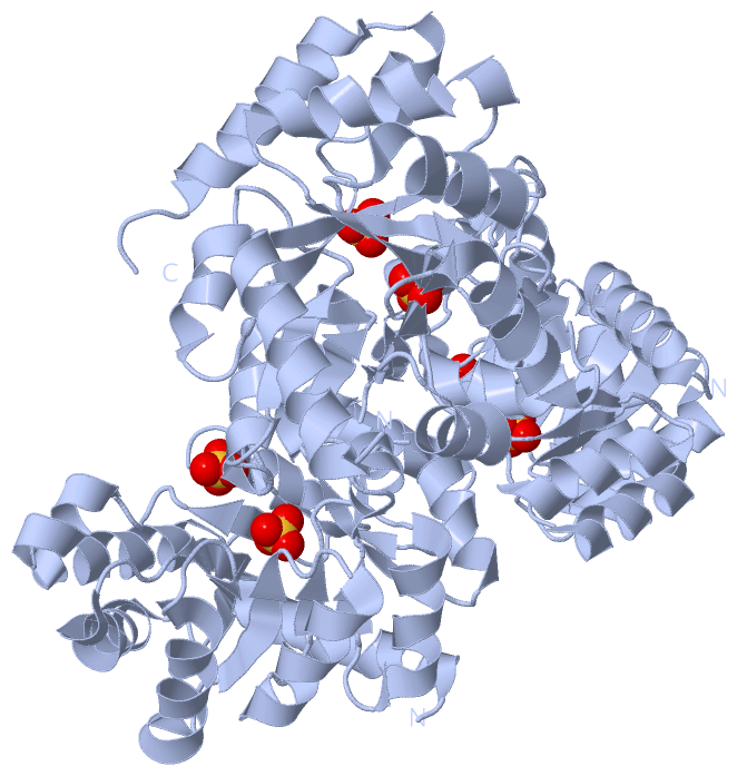
Asymmetric UnitChain A from PDB Type:PROTEIN Length:216 aligned with ALKD_PSEPU | P00885 from UniProtKB/Swiss-Prot Length:226 Alignment length:216 20 30 40 50 60 70 80 90 100 110 120 130 140 150 160 170 180 190 200 210 220 ALKD_PSEPU 11 LSMADKAARIDAICEKARILPVITIAREEDILPLADALAAGGIRTLEVTLRSQHGLKAIQVLREQRPELCVGAGTVLDRSMFAAVEAAGAQFVVTPGITEDILEAGVDSEIPLLPGISTPSEIMMGYALGYRRFKLFPAEISGGVAAIKAFGGPFGDIRFCPTGGVNPANVRNYMALPNVMCVGTGWMLDSSWIKNGDWARIEACSAEAIALLDAN 226 SCOP domains d1mxsa_ A: KDPG aldolase SCOP domains CATH domains 1mxsA00 A:11-226 Aldolase class I CATH domains Pfam domains ---------Aldolase-1mxsA01 A:20-215 ----------- Pfam domains SAPs(SNPs) ------------------------------------------------------------------------------------------------------------------------------------------------------------------------------------------------------------------------ SAPs(SNPs) PROSITE -----------------------------------------ALDOLASE_K------------------------------------------------------------------------------ALDOLASE_KDPG_------------------------------------------------------------------------- PROSITE Transcript ------------------------------------------------------------------------------------------------------------------------------------------------------------------------------------------------------------------------ Transcript 1mxs A 11 LSMADKAARIDAICEKARILPVITIAREEDILPLADALAAGGIRTLEVTLRSQHGLKAIQVLREQRPELCVGAGTVLDRSMFAAVEAAGAQFVVTPGITEDILEAGVDSEIPLLPGISTPSEIMMGYALGYRRFKLFPAEISGGVAAIKAFGGPFGDIRFCPTGGVNPANVRNYMALPNVMCVGTTWMLDSSWIKNGDWARIEACSAEAIALLDAN 226 20 30 40 50 60 70 80 90 100 110 120 130 140 150 160 170 180 190 200 210 220
|
||||||||||||||||||||
SCOP Domains (1, 1)
Asymmetric Unit
 |
CATH Domains (1, 1)
Asymmetric Unit
|
Pfam Domains (1, 1)| Asymmetric Unit |
Gene Ontology (4, 4)|
Asymmetric Unit(hide GO term definitions) Chain A (ALKD_PSEPU | P00885)
|
||||||||||||||||||||||||||||||||||||
Interactive Views
|
||||||||||||||||||||||||||||||||||||||||||||||||||||||||||||||||||||||||||||||||||||||||||||||||||||||||||||||||||||||||||||||||||||||||||||||||
Still Images
|
||||||||||||||||
Databases
|
||||||||||||||||||||||||||||||||||||||||||||||||||||||||||||||||||||||||||||||||||||||||||||||||||||||||||||||||||||||||||||||||||||||||||||||||||||||||||||||||
Analysis Tools
|
|||||||||||||||||||||||||||||||||||||||||||||||||||||||||||||
Entries Sharing at Least One Protein Chain (UniProt ID)
Related Entries Specified in the PDB File
|
|