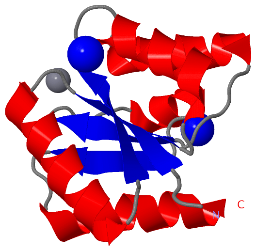
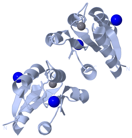
Show PDB file:   
         Plain Text   HTML   (compressed file size)
QuickSearch:   
by PDB,NDB,UniProt,PROSITE Code or Search Term(s)  
(-)Biol.Unit 1 - manually
(-)Asym.Unit - manually
(-)Asymmetric Unit
(-)Asym. Unit - sites
(-)Biological Unit 1
(-)Biol. Unit 1 - sites
collapse expand < >
Image Biol.Unit 1 - manually
Biol.Unit 1 - manually  (Jmol Viewer)
Image Asym.Unit - manually
Asym.Unit - manually  (Jmol Viewer)
Image Asymmetric Unit
Asymmetric Unit  (Jmol Viewer)
Image Asym. Unit - sites
Asym. Unit - sites  (Jmol Viewer)
Image Biological Unit 1
Biological Unit 1  (Jmol Viewer)
Image Biol. Unit 1 - sites
Biol. Unit 1 - sites  (Jmol Viewer)

(-) Description

Title :  CRYSTAL STRUCTURE OF THE PHOP RECEIVER DOMAIN FROM BACILLUS SUBTILIS
 
Authors :  C. Birck, Y. Chen, F. M. Hulett, J. P. Samama, Structural Proteomics In Europe (Spine)
Date :  26 Sep 02  (Deposition) - 16 Oct 02  (Release) - 24 Feb 09  (Revision)
Method :  X-RAY DIFFRACTION
Resolution :  1.60
Chains :  Asym. Unit :  A
Biol. Unit 1:  A  (2x)
Keywords :  Phosphate Regulon, Transcriptional Regulatory Protein, Alpha/Beta Doubly Wound Fold, Response Regulator, Phosphorylation, Asymmetric Interface, Tandem Association, Structural Proteomics In Europe, Spine, Structural Genomics (Keyword Search: [Gene Ontology, PubMed, Web (Google))
 
Reference :  C. Birck, Y. Chen, F. M. Hulett, J. P. Samama
The Crystal Structure Of The Phosphorylation Domain In Phop Reveals A Functional Tandem Association Mediated By An Asymmetric Interface
J. Bacteriol. V. 185 254 2003
PubMed-ID: 12486062  |  Reference-DOI: 10.1128/JB.185.1.254-261.2003
(for further references see the PDB file header)

(-) Compounds

Molecule 1 - PHOP RESPONSE REGULATOR
    ChainsA
    EngineeredYES
    Expression SystemESCHERICHIA COLI BL21(DE3)
    Expression System PlasmidPET16B
    Expression System StrainBL21(DE3)
    Expression System Taxid469008
    Expression System Vector TypePLASMID
    FragmentN-TERMINAL DOMAIN
    GenePHOP
    Organism ScientificBACILLUS SUBTILIS
    Organism Taxid1423
    SynonymALKALINE PHOSPHATASE SYNTHESIS TRANSCRIPTIONAL REGULATORY PROTEIN PHOP

 Structural Features

(-) Chains, Units

  1
Asymmetric Unit A
Biological Unit 1 (2x)A

Summary Information (see also Sequences/Alignments below)

(-) Ligands, Modified Residues, Ions  (2, 3)

Asymmetric Unit (2, 3)
No.NameCountTypeFull Name
1MN1Ligand/IonMANGANESE (II) ION
2NA2Ligand/IonSODIUM ION
Biological Unit 1 (0, 0)
No.NameCountTypeFull Name
1MN-1Ligand/IonMANGANESE (II) ION
2NA-1Ligand/IonSODIUM ION

(-) Sites  (3, 3)

Asymmetric Unit (3, 3)
No.NameEvidenceResiduesDescription
1AC1SOFTWAREASP A:10 , ASP A:53 , MET A:55 , HOH A:214 , HOH A:354 , HOH A:373BINDING SITE FOR RESIDUE MN A 200
2AC2SOFTWAREGLU A:37 , HOH A:355 , HOH A:356 , HOH A:364 , HOH A:372 , HOH A:375BINDING SITE FOR RESIDUE NA A 201
3AC3SOFTWAREPHE A:75 , GLY A:96 , ASP A:98 , HOH A:246 , HOH A:253BINDING SITE FOR RESIDUE NA A 202

(-) SS Bonds  (0, 0)

(no "SS Bond" information available for 1MVO)

(-) Cis Peptide Bonds  (1, 1)

Asymmetric Unit
No.Residues
1Lys A:103 -Pro A:104

 Sequence-Structure Mapping

(-) SAPs(SNPs)/Variants  (0, 0)

(no "SAP(SNP)/Variant" information available for 1MVO)

(-) PROSITE Motifs  (1, 1)

Asymmetric Unit (1, 1)
 PROSITEUniProtKBPDB
No.IDACDescriptionIDLocationCountLocation
1RESPONSE_REGULATORYPS50110 Response regulatory domain profile.PHOP_BACSU4-118  1A:4-118
Biological Unit 1 (1, 2)
 PROSITEUniProtKBPDB
No.IDACDescriptionIDLocationCountLocation
1RESPONSE_REGULATORYPS50110 Response regulatory domain profile.PHOP_BACSU4-118  2A:4-118

(-) Exons   (0, 0)

(no "Exon" information available for 1MVO)

(-) Sequences/Alignments

Asymmetric Unit
   Reformat: Number of residues per line =  ('0' or empty: single-line sequence representation)
  Number of residues per labelling interval =   
  UniProt sequence: complete  aligned part    
   Show mapping: SCOP domains CATH domains Pfam domains Secondary structure (by author)
SAPs(SNPs) PROSITE motifs Exons
(details for a mapped element are shown in a popup box when the mouse pointer rests over it)
Chain A from PDB  Type:PROTEIN  Length:121
 aligned with PHOP_BACSU | P13792 from UniProtKB/Swiss-Prot  Length:240

    Alignment length:121
                                    10        20        30        40        50        60        70        80        90       100       110       120 
           PHOP_BACSU     1 MNKKILVVDDEESIVTLLQYNLERSGYDVITASDGEEALKKAETEKPDLIVLDVMLPKLDGIEVCKQLRQQKLMFPILMLTAKDEEFDKVLGLELGADDYMTKPFSPREVNARVKAILRRS 121
               SCOP domains d1mvoa_ A: PhoP receiver domain                                                                                           SCOP domains
               CATH domains 1mvoA00 A:1-121  [code=3.40.50.2300, no name defined]                                                                     CATH domains
               Pfam domains ----Response_reg-1mvoA01 A:5-115                                                                                   ------ Pfam domains
         Sec.struct. author ...eeeee..hhhhhhhhhhhhhhh..eeeee.hhhhhhhhhhhhh..eeeee......hhhhhhhhhhhh.....eeeee......hhhhhhhh...eeee...hhhhhhhhhhhhhhh. Sec.struct. author
                 SAPs(SNPs) ------------------------------------------------------------------------------------------------------------------------- SAPs(SNPs)
                    PROSITE ---RESPONSE_REGULATORY  PDB: A:4-118 UniProt: 4-118                                                                   --- PROSITE
                 Transcript ------------------------------------------------------------------------------------------------------------------------- Transcript
                 1mvo A   1 MNKKILVVDDEESIVTLLQYNLERSGYDVITASDGEEALKKAETEKPDLIVLDVMLPKLDGIEVCKQLRQQKLMFPILMLTAKDEEFDKVLGLELGADDYMTKPFSPREVNARVKAILRRS 121
                                    10        20        30        40        50        60        70        80        90       100       110       120 

   Legend:   → Mismatch (orange background)
  - → Gap (green background, '-', border residues have a numbering label)
    → Modified Residue (blue background, lower-case, 'x' indicates undefined single-letter code, labelled with number + name)
  x → Chemical Group (purple background, 'x', labelled with number + name, e.g. ACE or NH2)
  extra numbering lines below/above indicate numbering irregularities and modified residue names etc., number ends below/above '|'

 Classification and Annotation

(-) SCOP Domains  (1, 1)

Asymmetric Unit

(-) CATH Domains  (1, 1)

Asymmetric Unit
(-)
Class: Alpha Beta (26913)

(-) Pfam Domains  (1, 1)

Asymmetric Unit
(-)
Clan: CheY (97)

(-) Gene Ontology  (7, 7)

Asymmetric Unit(hide GO term definitions)
Chain A   (PHOP_BACSU | P13792)
molecular function
    GO:0003677    DNA binding    Any molecular function by which a gene product interacts selectively and non-covalently with DNA (deoxyribonucleic acid).
biological process
    GO:0006817    phosphate ion transport    The directed movement of phosphate into, out of or within a cell, or between cells, by means of some agent such as a transporter or pore.
    GO:0000160    phosphorelay signal transduction system    A conserved series of molecular signals found in prokaryotes and eukaryotes; involves autophosphorylation of a histidine kinase and the transfer of the phosphate group to an aspartate that then acts as a phospho-donor to response regulator proteins.
    GO:0006355    regulation of transcription, DNA-templated    Any process that modulates the frequency, rate or extent of cellular DNA-templated transcription.
    GO:0006351    transcription, DNA-templated    The cellular synthesis of RNA on a template of DNA.
    GO:0006810    transport    The directed movement of substances (such as macromolecules, small molecules, ions) or cellular components (such as complexes and organelles) into, out of or within a cell, or between cells, or within a multicellular organism by means of some agent such as a transporter, pore or motor protein.
cellular component
    GO:0005737    cytoplasm    All of the contents of a cell excluding the plasma membrane and nucleus, but including other subcellular structures.

 Visualization

(-) Interactive Views

Asymmetric Unit
  Complete Structure
    Jena3D(integrated viewing of ligand, site, SAP, PROSITE, SCOP information)
    WebMol | AstexViewer[tm]@PDBe
(Java Applets, require no local installation except for Java; loading may be slow)
    STRAP
(Java WebStart application, automatic local installation, requires Java; full application with system access!)
    RasMol
(require local installation)
    Molscript (VRML)
(requires installation of a VRML viewer; select preferred view via VRML and generate a mono or stereo PDF format file)
 
  Ligands, Modified Residues, Ions
    MN  [ RasMol | Jena3D ]  +environment [ RasMol | Jena3D ]
    NA  [ RasMol | Jena3D ]  +environment [ RasMol | Jena3D ]
 
  Sites
    AC1  [ RasMol ]  +environment [ RasMol ]
    AC2  [ RasMol ]  +environment [ RasMol ]
    AC3  [ RasMol ]  +environment [ RasMol ]
 
  Cis Peptide Bonds
    Lys A:103 - Pro A:104   [ RasMol ]  
 
Biological Unit
  Complete Structure
    Biological Unit 1  [ Jena3D ]

(-) Still Images

Jmol
  protein: cartoon or spacefill or dots and stick; nucleic acid: cartoon and stick; ligands: spacefill; active site: stick
Molscript
  protein, nucleic acid: cartoon; ligands: spacefill; active site: ball and stick

 Databases and Analysis Tools

(-) Databases

Access by PDB/NDB ID
  1mvo
    Family and Domain InformationProDom | SYSTERS
    General Structural InformationGlycoscienceDB | MMDB | NDB | OCA | PDB | PDBe | PDBj | PDBsum | PDBWiki | PQS | PROTEOPEDIA
    Orientation in MembranesOPM
    Protein SurfaceSURFACE
    Secondary StructureDSSP (structure derived) | HSSP (homology derived)
    Structural GenomicsGeneCensus
    Structural NeighboursCE | VAST
    Structure ClassificationCATH | Dali | SCOP
    Validation and Original DataBMRB Data View | BMRB Restraints Grid | EDS | PROCHECK | RECOORD | WHAT_CHECK
 
Access by UniProt ID/Accession number
  PHOP_BACSU | P13792
    Comparative Protein Structure ModelsModBase
    Genomic InformationEnsembl
    Protein-protein InteractionDIP
    Sequence, Family and Domain InformationInterPro | Pfam | SMART | UniProtKB/SwissProt
 
Access by Enzyme Classificator   (EC Number)
  (no 'Enzyme Classificator' available)
    General Enzyme InformationBRENDA | EC-PDB | Enzyme | IntEnz
    PathwayKEGG | MetaCyc
 
Access by Disease Identifier   (MIM ID)
  (no 'MIM ID' available)
    Disease InformationOMIM
 
Access by GenAge ID
  (no 'GenAge ID' available)
    Age Related InformationGenAge

(-) Analysis Tools

Access by PDB/NDB ID
    Domain InformationXDom
    Interatomic Contacts of Structural UnitsCSU
    Ligand-protein ContactsLPC
    Protein CavitiescastP
    Sequence and Secondary StructurePDBCartoon
    Structure AlignmentSTRAP(Java WebStart application, automatic local installation, requires Java; full application with system access!)
    Structure and Sequence BrowserSTING
 
Access by UniProt ID/Accession number
  PHOP_BACSU | P13792
    Protein Disorder PredictionDisEMBL | FoldIndex | GLOBPLOT (for more information see DisProt)

 Related Entries

(-) Entries Sharing at Least One Protein Chain (UniProt ID)

(no "Entries Sharing at Least One Protein Chain" available for 1MVO)

(-) Related Entries Specified in the PDB File

(no "Related Entries Specified in the PDB File" available for 1MVO)