|
|
|
|
 Description
|
|
Compounds
|
||||||||||||||||||||||||||||||||||||||||||||||||||||
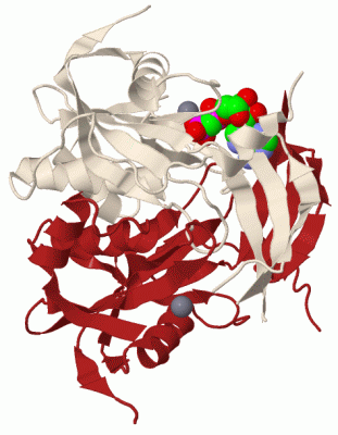
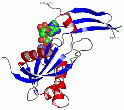
Chains, Units
Summary Information (see also Sequences/Alignments below) |
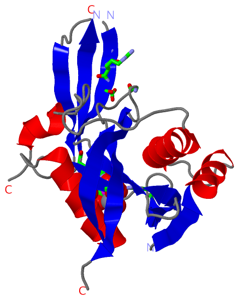
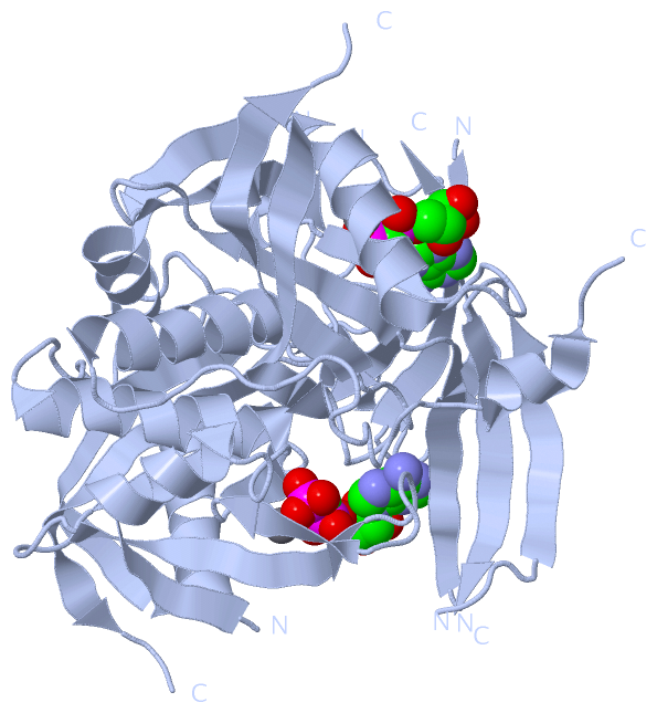
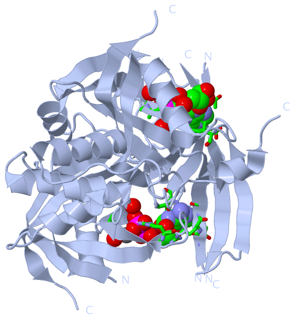
Ligands, Modified Residues, Ions (2, 2)| Asymmetric Unit (2, 2) Biological Unit 1 (1, 2) |
Sites (2, 2)
Asymmetric Unit (2, 2)
|
SS Bonds (0, 0)| (no "SS Bond" information available for 1MR2) |
Cis Peptide Bonds (0, 0)| (no "Cis Peptide Bond" information available for 1MR2) |
SAPs(SNPs)/Variants (0, 0)| (no "SAP(SNP)/Variant" information available for 1MR2) |
PROSITE Motifs (0, 0)| (no "PROSITE Motif" information available for 1MR2) |
Exons (0, 0)| (no "Exon" information available for 1MR2) |
Sequences/Alignments
Asymmetric UnitChain A from PDB Type:PROTEIN Length:187 aligned with O33199_MYCTO | O33199 from UniProtKB/TrEMBL Length:207 Alignment length:202 15 25 35 45 55 65 75 85 95 105 115 125 135 145 155 165 175 185 195 205 O33199_MYCTO 6 FETISSETLHTGAIFALRRDQVRMPGGGIVTREVVEHFGAVAIVAMDDNGNIPMVYQYRHTYGRRLWELPAGLLDVAGEPPHLTAARELREEVGLQASTWQVLVDLDTAPGFSDESVRVYLATGLREVGRPEAHHEEADMTMGWYPIAEAARRVLRGEIVNSIAIAGVLAVHAVTTGFAQPRPLDTEWIDRPTAFAARRAER 207 SCOP domains d1mr2a_ A: ADP-ribose p yrophosphatase SCOP domains CATH domains 1mr2A00 A:6-207 Nucleos ide Triphosphate Pyrophosphohydrolase CATH domains Pfam domains ---------------------------------------------------------------------------------------------------------------------------------------------------------------------------------------------------------- Pfam domains SAPs(SNPs) ---------------------------------------------------------------------------------------------------------------------------------------------------------------------------------------------------------- SAPs(SNPs) PROSITE ---------------------------------------------------------------------------------------------------------------------------------------------------------------------------------------------------------- PROSITE Transcript ---------------------------------------------------------------------------------------------------------------------------------------------------------------------------------------------------------- Transcript 1mr2 A 6 FETISSETLHTGAIFALRRDQVR-----IVTREVVEHFGAVAIVAMDDNGNIPMVYQYRHTYGRRLWELPAGLLDVAGEPPHLTAARELREEVGLQASTWQVLVDLDTAPGFSDESVRVYLATGLREVGR----------TMGWYPIAEAARRVLRGEIVNSIAIAGVLAVHAVTTGFAQPRPLDTEWIDRPTAFAARRAER 207 15 25 | 35 45 55 65 75 85 95 105 115 125 135 -| 155 165 175 185 195 205 28 34 135 146
|
||||||||||||||||||||
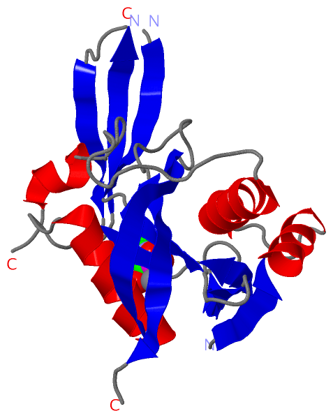
SCOP Domains (1, 1)
Asymmetric Unit
 |
CATH Domains (1, 1)
Asymmetric Unit
|
Pfam Domains (0, 0)| (no "Pfam Domain" information available for 1MR2) |
Gene Ontology (3, 3)|
Asymmetric Unit(hide GO term definitions) Chain A (O33199_MYCTO | O33199)
|
||||||||||||||||||||||||
Interactive Views
|
||||||||||||||||||||||||||||||||||||||||||||||||||||||||||||||||||||||||||||||||||||||||||||||||||||||||||||||||||||||||||||||||||||||||||||||||||||||
Still Images
|
||||||||||||||||
Databases
|
||||||||||||||||||||||||||||||||||||||||||||||||||||||||||||||||||||||||||||||||||||||||||||||||||||||||||||||||||||||||||||||||||||||||||||||||||||||||||||||||
Analysis Tools
|
|||||||||||||||||||||||||||||||||||||||||||||||||||||||||||||
Entries Sharing at Least One Protein Chain (UniProt ID)
Related Entries Specified in the PDB File
|
|