|
|
|
|
 Description
|
|
Compounds
|
||||||||||||||||||||||||||||||||||||||||||||
Chains, Units
Summary Information (see also Sequences/Alignments below) |
Ligands, Modified Residues, Ions (1, 2)
Asymmetric Unit (1, 2)
|
Sites (2, 2)
Asymmetric Unit (2, 2)
|
SS Bonds (0, 0)| (no "SS Bond" information available for 1MQV) |
Cis Peptide Bonds (0, 0)| (no "Cis Peptide Bond" information available for 1MQV) |
SAPs(SNPs)/Variants (0, 0)| (no "SAP(SNP)/Variant" information available for 1MQV) |
PROSITE Motifs (1, 2)
Asymmetric Unit (1, 2)
|
||||||||||||||||||||||||||||||||||||||||||||||||||||||||||||||||||||||||
Exons (0, 0)| (no "Exon" information available for 1MQV) |
Sequences/Alignments
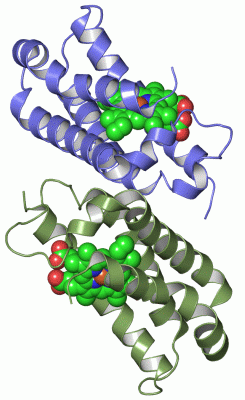
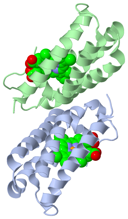
Asymmetric UnitChain A from PDB Type:PROTEIN Length:123 aligned with CYCP_RHOPA | P00149 from UniProtKB/Swiss-Prot Length:146 Alignment length:123 31 41 51 61 71 81 91 101 111 121 131 141 CYCP_RHOPA 22 QTDVIAQRKAILKQMGEATKPIAAMLKGEAKFDQAVVQKSLAAIADDSKKLPALFPADSKTGGDTAALPKIWEDKAKFDDLFAKLAAAATAAQGTIKDEASLKANIGGVLGNCKSCHDDFRAK 144 SCOP domains d1mqva_ A: Cytochrome c' SCOP domains CATH domains 1mqvA00 A:1-123 [code=1.20.120.10, no name defined] CATH domains Pfam domains --------------------------------------------------------------------------------------------------------------------------- Pfam domains SAPs(SNPs) --------------------------------------------------------------------------------------------------------------------------- SAPs(SNPs) PROSITE --CYTCII PDB: A:3-122 UniProt: 24-143 - PROSITE Transcript --------------------------------------------------------------------------------------------------------------------------- Transcript 1mqv A 1 ATDVIAQRKAILKQMGEATKPIAAMLKGEAKWDQAVVQKSLAAIADDSKKLPALFPADSKTGGDTAALPKIFEDKAKFDDLFAKLAAAATAAQGTIKDEASLKANIGGVLGNCKSCHDDFRAK 123 10 20 30 40 50 60 70 80 90 100 110 120 Chain B from PDB Type:PROTEIN Length:123 aligned with CYCP_RHOPA | P00149 from UniProtKB/Swiss-Prot Length:146 Alignment length:123 31 41 51 61 71 81 91 101 111 121 131 141 CYCP_RHOPA 22 QTDVIAQRKAILKQMGEATKPIAAMLKGEAKFDQAVVQKSLAAIADDSKKLPALFPADSKTGGDTAALPKIWEDKAKFDDLFAKLAAAATAAQGTIKDEASLKANIGGVLGNCKSCHDDFRAK 144 SCOP domains d1mqvb_ B: Cytochrome c' SCOP domains CATH domains 1mqvB00 B:1-123 [code=1.20.120.10, no name defined] CATH domains Pfam domains (1) -Cytochrom_C_2-1mqvB01 B:2-121 -- Pfam domains (1) Pfam domains (2) -Cytochrom_C_2-1mqvB02 B:2-121 -- Pfam domains (2) SAPs(SNPs) --------------------------------------------------------------------------------------------------------------------------- SAPs(SNPs) PROSITE --CYTCII PDB: B:3-122 UniProt: 24-143 - PROSITE Transcript --------------------------------------------------------------------------------------------------------------------------- Transcript 1mqv B 1 ATDVIAQRKAILKQMGEATKPIAAMLKGEAKWDQAVVQKSLAAIADDSKKLPALFPADSKTGGDTAALPKIFEDKAKFDDLFAKLAAAATAAQGTIKDEASLKANIGGVLGNCKSCHDDFRAK 123 10 20 30 40 50 60 70 80 90 100 110 120
|
||||||||||||||||||||
SCOP Domains (1, 2)
Asymmetric Unit
 |
CATH Domains (1, 2)
Asymmetric Unit
|
Pfam Domains (1, 2)
Asymmetric Unit
|
Gene Ontology (7, 7)|
Asymmetric Unit(hide GO term definitions) Chain A,B (CYCP_RHOPA | P00149)
|
||||||||||||||||||||||||||||||||||||||||||||||||||||||||||||
Interactive Views
|
||||||||||||||||||||||||||||||||||||||||||||||||||||||||||||||||||||||||||||||||||||||||||||||||||||||||||||||||||||||||||||||||||||||||||||||||||||
Still Images
|
||||||||||||||||
Databases
|
||||||||||||||||||||||||||||||||||||||||||||||||||||||||||||||||||||||||||||||||||||||||||||||||||||||||||||||||||||||||||||||||||||||||||||||||||||||||||||||||
Analysis Tools
|
|||||||||||||||||||||||||||||||||||||||||||||||||||||||||||||
Entries Sharing at Least One Protein Chain (UniProt ID)
Related Entries Specified in the PDB File
|
|