Show PDB file:   
         Plain Text   HTML   (compressed file size)
QuickSearch:   
by PDB,NDB,UniProt,PROSITE Code or Search Term(s)  
(-)NMR Structure - manually
(-)NMR Structure - model 1
(-)NMR Structure - all models
collapse expand < >
Image NMR Structure - manually
NMR Structure - manually  (Jmol Viewer)
Image NMR Structure - model 1
NMR Structure - model 1  (Jmol Viewer)
Image NMR Structure - all models
NMR Structure - all models  (Jmol Viewer)

(-) Description

Title :  RECOMBINANT CUCURBITA MAXIMA TRYPSIN INHIBITOR V (RCMTI-V) (NMR, MINIMIZED AVERAGE STRUCTURE)
 
Authors :  M. Cai, Y. Gong, Y. Huang, J. Liu, O. Prakash, L. Wen, J. J. Wen, J. - K. Huang, R. Krishnamoorthi
Date :  26 Oct 95  (Deposition) - 03 Apr 96  (Release) - 24 Feb 09  (Revision)
Method :  SOLUTION NMR
Resolution :  NOT APPLICABLE
Chains :  NMR Structure  :  A
Keywords :  Serine Protease Inhibitor (Rcmti-V) (Keyword Search: [Gene Ontology, PubMed, Web (Google))
 
Reference :  J. Liu, O. Prakash, M. Cai, Y. Gong, Y. Huang, L. Wen, J. J. Wen, J. K. Huang, R. Krishnamoorthi
Solution Structure And Backbone Dynamics Of Recombinant Cucurbita Maxima Trypsin Inhibitor-V Determined By Nmr Spectroscopy.
Biochemistry V. 35 1516 1996
PubMed-ID: 8634282  |  Reference-DOI: 10.1021/BI952466D
(for further references see the PDB file header)

(-) Compounds

Molecule 1 - TRYPSIN INHIBITOR V
    ChainsA
    EngineeredYES
    Expression SystemESCHERICHIA COLI
    Expression System Taxid562
    OrganSEED
    Organism CommonWINTER SQUASH
    Organism ScientificCUCURBITA MAXIMA
    Organism Taxid3661
    SynonymRCMTI-V

 Structural Features

(-) Chains, Units

  
NMR Structure 

Summary Information (see also Sequences/Alignments below)

(-) Ligands, Modified Residues, Ions  (0, 0)

(no "Ligand,Modified Residues,Ions" information available for 1MIT)

(-) Sites  (0, 0)

(no "Site" information available for 1MIT)
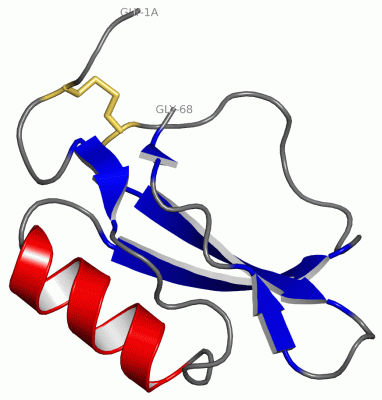
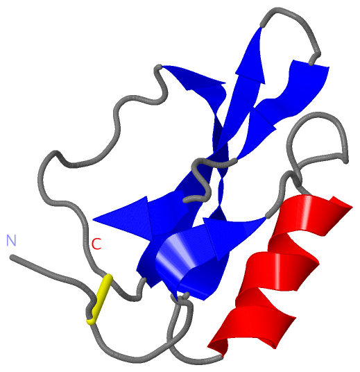
(-) SS Bonds  (1, 1)

NMR Structure
No.Residues
1A:3 -A:48

(-) Cis Peptide Bonds  (0, 0)

(no "Cis Peptide Bond" information available for 1MIT)

 Sequence-Structure Mapping

(-) SAPs(SNPs)/Variants  (0, 0)

(no "SAP(SNP)/Variant" information available for 1MIT)

(-) PROSITE Motifs  (1, 1)

NMR Structure (1, 1)
 PROSITEUniProtKBPDB
No.IDACDescriptionIDLocationCountLocation
1POTATO_INHIBITORPS00285 Potato inhibitor I family signature.ITH5_CUCMA9-20  1A:9-20

(-) Exons   (0, 0)

(no "Exon" information available for 1MIT)

(-) Sequences/Alignments

NMR Structure
   Reformat: Number of residues per line =  ('0' or empty: single-line sequence representation)
  Number of residues per labelling interval =   
  UniProt sequence: complete  aligned part    
   Show mapping: SCOP domains CATH domains Pfam domains Secondary structure (by author)
SAPs(SNPs) PROSITE motifs Exons
(details for a mapped element are shown in a popup box when the mouse pointer rests over it)
Chain A from PDB  Type:PROTEIN  Length:69
 aligned with ITH5_CUCMA | P19873 from UniProtKB/Swiss-Prot  Length:68

    Alignment length:69
                             1                                                                   
                             |       9        19        29        39        49        59         
            ITH5_CUCMA    - -SSCPGKSSWPHLVGVGGSVAKAIIERQNPNVKAVILEEGTPVTKDFRCNRVRIWVNKRGLVVSPPRIG 68
               SCOP domains d1mita_ A: Trypsin inhibitor V                                        SCOP domains
               CATH domains 1mitA00 A:1A-68 Trypsin Inhibitor V, subunit A                        CATH domains
               Pfam domains ------potato_inhibit-1mitA01 A:6-68                                   Pfam domains
         Sec.struct. author .......eee........hhhhhhhhhhh...eeeeeee...........eeeeeee...eee...ee. Sec.struct. author
                 SAPs(SNPs) --------------------------------------------------------------------- SAPs(SNPs)
                    PROSITE ---------POTATO_INHIB------------------------------------------------ PROSITE
                 Transcript --------------------------------------------------------------------- Transcript
                  1mit A 1A GSSCPGKSSWPHLVGVGGSVAKAIIERQNPNVKAVILEEGTPVTKDFRCNRVRIWVNKRGLVVSPPRIG 68
                            |        9        19        29        39        49        59         
                            |                                                                    
                           1A                                                                    

   Legend:   → Mismatch (orange background)
  - → Gap (green background, '-', border residues have a numbering label)
    → Modified Residue (blue background, lower-case, 'x' indicates undefined single-letter code, labelled with number + name)
  x → Chemical Group (purple background, 'x', labelled with number + name, e.g. ACE or NH2)
  extra numbering lines below/above indicate numbering irregularities and modified residue names etc., number ends below/above '|'

 Classification and Annotation

(-) SCOP Domains  (1, 1)

NMR Structure

(-) CATH Domains  (1, 1)

NMR Structure
(-)
Class: Alpha Beta (26913)

(-) Pfam Domains  (1, 1)

NMR Structure
(-)
Clan: CI-2 (32)

(-) Gene Ontology  (5, 5)

NMR Structure(hide GO term definitions)
Chain A   (ITH5_CUCMA | P19873)
molecular function
    GO:0030414    peptidase inhibitor activity    Stops, prevents or reduces the activity of a peptidase, any enzyme that catalyzes the hydrolysis peptide bonds.
    GO:0004867    serine-type endopeptidase inhibitor activity    Stops, prevents or reduces the activity of serine-type endopeptidases, enzymes that catalyze the hydrolysis of nonterminal peptide bonds in a polypeptide chain; a serine residue (and a histidine residue) are at the active center of the enzyme.
biological process
    GO:0010951    negative regulation of endopeptidase activity    Any process that decreases the frequency, rate or extent of endopeptidase activity, the endohydrolysis of peptide bonds within proteins.
    GO:0010466    negative regulation of peptidase activity    Any process that stops or reduces the rate of peptidase activity, the hydrolysis of peptide bonds within proteins.
    GO:0009611    response to wounding    Any process that results in a change in state or activity of a cell or an organism (in terms of movement, secretion, enzyme production, gene expression, etc.) as a result of a stimulus indicating damage to the organism.

 Visualization

(-) Interactive Views

NMR Structure
  Complete Structure
    Jena3D(integrated viewing of ligand, site, SAP, PROSITE, SCOP information)
    WebMol | AstexViewer[tm]@PDBe
(Java Applets, require no local installation except for Java; loading may be slow)
    STRAP
(Java WebStart application, automatic local installation, requires Java; full application with system access!)
    RasMol
(require local installation)
    Molscript (VRML)
(requires installation of a VRML viewer; select preferred view via VRML and generate a mono or stereo PDF format file)
 
  Ligands, Modified Residues, Ions
(no "Ligands, Modified Residues, Ions" information available for 1mit)
 
  Sites
(no "Sites" information available for 1mit)
 
  Cis Peptide Bonds
(no "Cis Peptide Bonds" information available for 1mit)
 

(-) Still Images

Jmol
  protein: cartoon or spacefill or dots and stick; nucleic acid: cartoon and stick; ligands: spacefill; active site: stick
Molscript
  protein, nucleic acid: cartoon; ligands: spacefill; active site: ball and stick

 Databases and Analysis Tools

(-) Databases

Access by PDB/NDB ID
  1mit
    Family and Domain InformationProDom | SYSTERS
    General Structural InformationGlycoscienceDB | MMDB | NDB | OCA | PDB | PDBe | PDBj | PDBsum | PDBWiki | PQS | PROTEOPEDIA
    Orientation in MembranesOPM
    Protein SurfaceSURFACE
    Secondary StructureDSSP (structure derived) | HSSP (homology derived)
    Structural GenomicsGeneCensus
    Structural NeighboursCE | VAST
    Structure ClassificationCATH | Dali | SCOP
    Validation and Original DataBMRB Data View | BMRB Restraints Grid | EDS | PROCHECK | RECOORD | WHAT_CHECK
 
Access by UniProt ID/Accession number
  ITH5_CUCMA | P19873
    Comparative Protein Structure ModelsModBase
    Genomic InformationEnsembl
    Protein-protein InteractionDIP
    Sequence, Family and Domain InformationInterPro | Pfam | SMART | UniProtKB/SwissProt
 
Access by Enzyme Classificator   (EC Number)
  (no 'Enzyme Classificator' available)
    General Enzyme InformationBRENDA | EC-PDB | Enzyme | IntEnz
    PathwayKEGG | MetaCyc
 
Access by Disease Identifier   (MIM ID)
  (no 'MIM ID' available)
    Disease InformationOMIM
 
Access by GenAge ID
  (no 'GenAge ID' available)
    Age Related InformationGenAge

(-) Analysis Tools

Access by PDB/NDB ID
    Domain InformationXDom
    Interatomic Contacts of Structural UnitsCSU
    Ligand-protein ContactsLPC
    Protein CavitiescastP
    Sequence and Secondary StructurePDBCartoon
    Structure AlignmentSTRAP(Java WebStart application, automatic local installation, requires Java; full application with system access!)
    Structure and Sequence BrowserSTING
 
Access by UniProt ID/Accession number
  ITH5_CUCMA | P19873
    Protein Disorder PredictionDisEMBL | FoldIndex | GLOBPLOT (for more information see DisProt)

 Related Entries

(-) Entries Sharing at Least One Protein Chain (UniProt ID)

UniProtKB/Swiss-Prot
        ITH5_CUCMA | P198731hym 1tin

(-) Related Entries Specified in the PDB File

(no "Related Entries Specified in the PDB File" available for 1MIT)