|
|
|
|
 Description
|
|
Compounds
|
||||||||||||||||||||
Chains, Units
Summary Information (see also Sequences/Alignments below) |
Ligands, Modified Residues, Ions (4, 24)
Asymmetric Unit (4, 24)
|
Sites (4, 4)
Asymmetric Unit (4, 4)
|
SS Bonds (0, 0)| (no "SS Bond" information available for 1M24) |
Cis Peptide Bonds (0, 0)| (no "Cis Peptide Bond" information available for 1M24) |
SAPs(SNPs)/Variants (0, 0)| (no "SAP(SNP)/Variant" information available for 1M24) |
PROSITE Motifs (0, 0)| (no "PROSITE Motif" information available for 1M24) |
Exons (0, 0)| (no "Exon" information available for 1M24) |
Sequences/Alignments
Asymmetric Unit
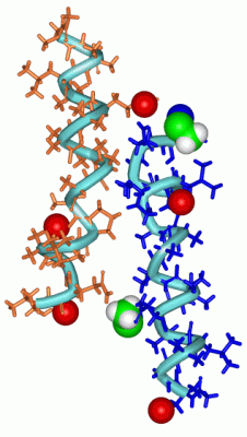
Chain A from PDB Type:PROTEIN Length:19
SCOP domains d1m24a_ A: SCOP domains
CATH domains ------------------- CATH domains
Pfam domains ------------------- Pfam domains
SAPs(SNPs) ------------------- SAPs(SNPs)
PROSITE ------------------- PROSITE
Transcript ------------------- Transcript
1m24 A 0 xxGxLxQxxxAAxPLxxQx 18
|| | | ||9 | || |
|| | | ||| | 16-AIB
0-ACE| ||| | | 18-VOL
1-AIB ||| | |
3-AIB|| | |
5-AIB | |
7-AIB| |
8-AIB |
9-AIB |
12-AIB
Chain B from PDB Type:PROTEIN Length:19
SCOP domains d1m24b_ B: SCOP domains
CATH domains ------------------- CATH domains
Pfam domains ------------------- Pfam domains
SAPs(SNPs) ------------------- SAPs(SNPs)
PROSITE ------------------- PROSITE
Transcript ------------------- Transcript
1m24 B 0 xxGxLxQxxxAAxPLxxQx 18
|| | | ||9 | || |
|| | | ||| | 16-AIB
0-ACE| ||| | | 18-VOL
1-AIB ||| | |
3-AIB|| | |
5-AIB | |
7-AIB| |
8-AIB |
9-AIB |
12-AIB
|
||||||||||||||||||||
SCOP Domains (1, 2)
Asymmetric Unit
|
CATH Domains (0, 0)| (no "CATH Domain" information available for 1M24) |
Pfam Domains (0, 0)| (no "Pfam Domain" information available for 1M24) |
Gene Ontology (0, 0)|
Asymmetric Unit(hide GO term definitions)
(no "Gene Ontology" information available for 1M24)
|
Interactive Views
|
|||||||||||||||||||||||||||||||||||||||||||||||||||||||||||||||||||||||||||||||||||||||||||||||||||||||||||||||||||||||||||||||||||||||||||||||||||||||||||||||||||||||||||||||||||||||
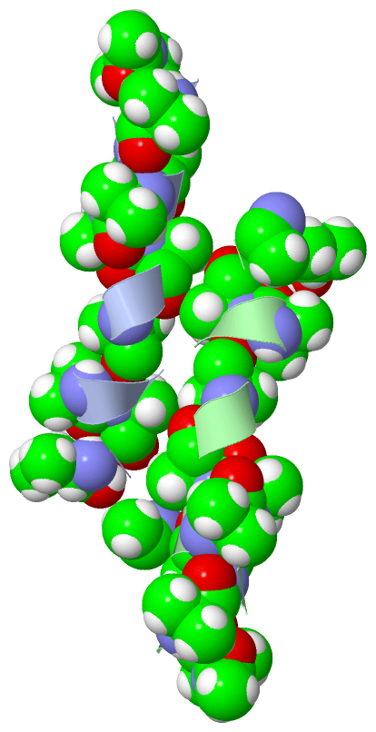
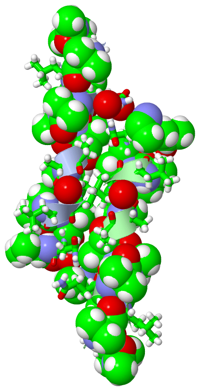
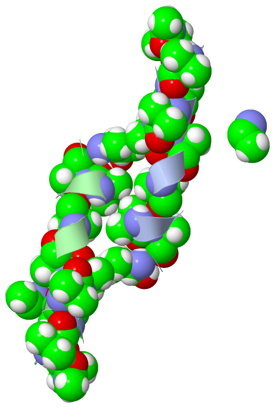
Still Images
|
||||||||||||||||||||||||||
Databases
|
||||||||||||||||||||||||||||||||||||||||||||||||||||||||||||||||||||||||||||||||||||||||||||||||||||||||||||||||||||||||||||||||||||||||||||||||||||||||||||||||
Analysis Tools
|
|||||||||||||||||||||||||||||||||||||||||||||||||||||||||||||
Entries Sharing at Least One Protein Chain (UniProt ID)
Related Entries Specified in the PDB File
|
|